3D Imaging Offers Insights on Lung Involvement in COVID-19
By MedImaging International staff writers
Posted on 01 Sep 2020
A novel three-dimensional (3D) imaging technique enables high resolution representation of damaged lung tissue following severe Covid-19. Posted on 01 Sep 2020
The new technique, developed at the University of Göttingen (Germany) and the Medical University of Hannover (MHH; Germany), is based on multi-scale phase contrast x-ray tomography, allows virtual histology and histopathology autopsy of the parenchymal architecture of unstained lung tissue from patients who succumbed to Covid-19. By combining parallel and cone beam geometry, autopsy samples with a maximum cross section of 4mm can be scanned and reconstructed at a resolution and image quality which allows for the segmentation of individual cells.

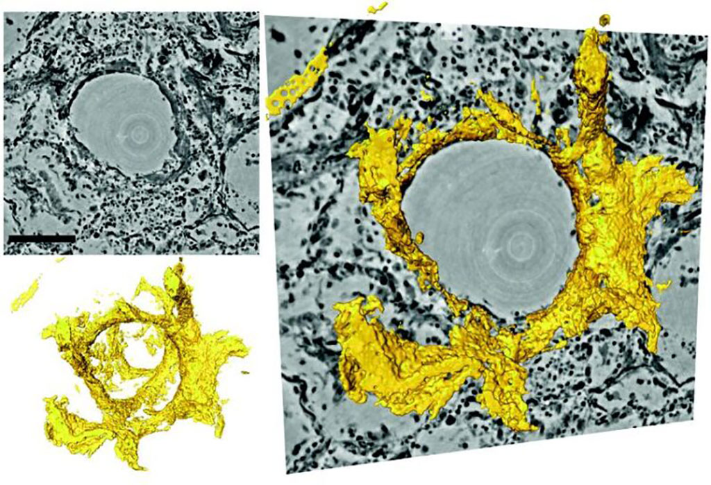
Image: 3D reconstruction around a pulmonary alveolus with hyaline membrane (yellow) (Photo courtesy of Tim Salditt/ University of Göttingen)
By using the zoom capability of the cone beam geometry, regions-of-interest are reconstructed with a minimum voxel size of 167 nm. In a proof-of-concept study, the researchers showed the capabilities of the new approach by visualizing diffuse alveolar damage (DAD), including prominent hyaline membrane formation, via mapping the 3D distribution and density of lymphocytes infiltrating the tissue, and by providing histograms of characteristic distances from tissue interior to the closest air compartment. The study was published on August 20, 2020, in eLife.
“Using zoom tomography, large areas of lung tissue embedded in wax can be scanned, enabling detailed examination to locate particularly interesting areas around inflammation, blood vessels, or bronchial tubes,” said lead author Professor Tim Salditt, PhD, of the University of Göttingen Institute of X-ray Physics. “Since X-rays penetrate deep into tissue, this enables scientists to understand the relation between the microscopic tissue structure and the larger functional architecture of an organ. This is important, for example, to visualize the tree of blood vessels down to the smallest capillaries.”
3D imaging via computerized tomography (CT) is not sufficient to detect tissue structure at cellular or sub-cellular resolution. The researchers therefore used phase contrast tomography, which exploits the different propagation velocities of X-rays in tissue to generate an intensity pattern. Special illumination optics and algorithms were then used to reconstruct sharp images from these patterns. This allowed examination of lung tissue at scalable size and resolution, yielding both larger overviews and close-up reconstructions.
Related Links:
University of Göttingen
Medical University of Hannover


