Imaging and AI Aid Intraoperative Brain Tumor Diagnosis
By MedImaging International staff writers
Posted on 15 Jan 2020
A workflow that combines advanced optical imaging with an artificial intelligence (AI) algorithm may accurately diagnose brain tumors in real time in the operating room, according to a new study.Posted on 15 Jan 2020
Developed at the University of California, San Francisco (UCSF; USA), the University of Michigan (U-M; Ann Arbor, USA), Columbia University (New York, NY, USA), and other institutions, the novel parallel workflow combines stimulated Raman histology (SRH, a label-free optical imaging method), and deep convolutional neural networks (CNNs) to predict diagnosis in near real time in an automated fashion. The CNNs, which were trained on over 2.5 million SRH images, built a hierarchy of recognizable histologic feature representations to help classify the major histopathologic classes of brain tumors.

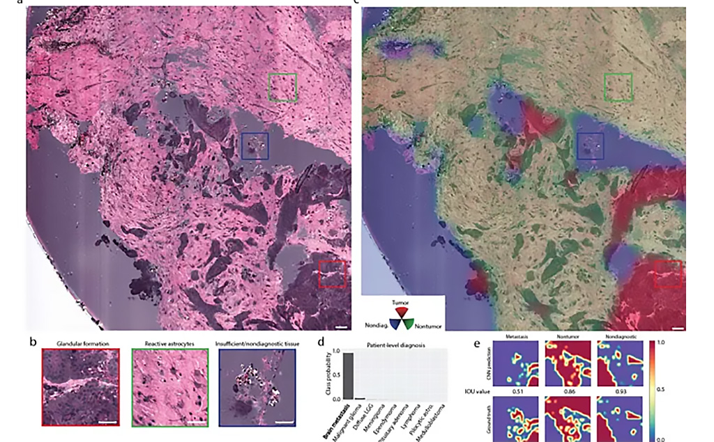
Image: Localization of metastatic brain tumor infiltration in SRH images (Photo courtesy of MGH)
The new workflow can diagnose brain tumors in less than 150 seconds, an order of magnitude faster than conventional histology techniques, which take 20–30 minutes. The authors also prospectively tested the workflow in a clinical trial of 278 patients with brain tumors, which demonstrated that the accuracy of CNN-based diagnosis of SRH images (94.6%) was slightly higher than pathologist-based interpretation of conventional histologic images (93.9%). The study was published on January 6, 2020, in Nature Medicine.
"As surgeons, we're limited to acting on what we can see; this technology allows us to see what would otherwise be invisible, to improve speed and accuracy in the operating room, and reduce the risk of misdiagnosis,” concluded lead author Todd Hollon, MD, of U-M, and colleagues. “Intraoperative cancer diagnosis can be streamlined, creating a complementary pathway for tissue diagnosis independent of a traditional pathology laboratory. With this imaging technology, cancer operations are safer and more effective than ever before.”
Related Links:
University of California, San Francisco
University of Michigan
Columbia University


