CC Imaging Capsule Requires No Colon Preparation
By MedImaging International staff writers
Posted on 14 Jan 2020
A new preparation-free ingestible scanning capsule helps prevent colorectal cancer (CC) through the detection of precancerous polyps.Posted on 14 Jan 2020
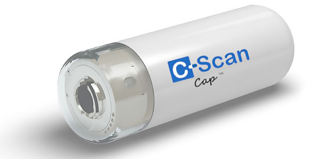
The Check-Cap (Isfiya, Israel) C-Scan System is comprised of an ultra-low dose X-ray capsule, an integrated positioning, control, and recording system, and proprietary software that is used to generate a three dimensional (3D) map of the inner lining of the colon. The heart of the system is the C-Scan Cap, an ingestible imaging capsule consisting of X-ray source that is collimated and rotated, forming three beams that scan the colon from within. Radiofrequency (RF) communication transmits the data to C-Scan Track, a tracking control and data collection unit comprised of three external patches that are worn on the patient's back.

Image: The Check-Cap prepless disposable capsule (Photo courtesy of Check-Cap)
C-Scan Track consists of an integrated positioning, control, and recording system that continuously tracks the capsule’s position and orientation along the colon, activates the capsule's scanning function during movement in the colon, and records and stores the capsule's information for future download. As the patient continues normal daily activities, the capsule is propelled through the gastrointestinal tract by natural motility, while continuously measuring the internal circumferential dimensions and tracking the position and orientation of the capsule within the body. The information is later used by C-Scan View to construct 2D and 3D images of the colon surface.
Patients swallow the capsule together with one tablespoon of radiopaque contrast solution; no fasting or bowel preparation is required. When C-Scan Cap has completed its passage, it is naturally excreted from the patient's body. The X-ray dose to which patients are exposed to for the entire procedure, from ingestion to excretion of the capsule, is similar to that of a single chest radiograph, significantly lower than conventional medical imaging procedures using X-rays such as computed tomography (CT) of the abdomen and pelvis and screening mammography.
“Check-Cap's prep-free, non-invasive technology meets the real need for colon cancer screening that's easy for patients. Patients are often hesitant to undergo colonoscopy due to the preparation, sedation, general discomfort and potential risks,” said Professor Oscar Lebwohl, MD, of Columbia University (New York, NY, USA). “If further studies demonstrate that Check-Cap is an accurate screening modality for colon cancer and polyps, then the Check-Cap system will be a viable testing alternative, allowing the screening of patients in greater numbers and the use of fewer resources compared with CTC and Colonoscopy.”
Related Links:
Check-Cap


