Miniature NMR Implant Measures Neuronal Activity
By MedImaging International staff writers
Posted on 17 Dec 2019
A highly sensitive nuclear magnetic resonance (NMR) implant probe enables brain physiology studies with enhanced spatial and temporal resolution.Posted on 17 Dec 2019
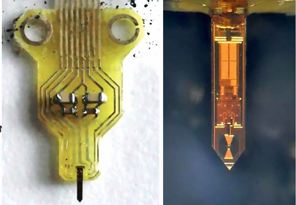
Developed at the Max-Planck-Institute for Biological Cybernetics (MPG; Tübingen, Germany), the University of Tübingen (Germany), and the University of Stuttgart (Germany), the capillary monolithic probe combines an ultra-sensitive 300 µm coil with a complete NMR transceiver, enabling in vivo measurements of blood oxygenation and flow in nanoliter volumes at a sampling rate of 200 Hz. To minimize the risk of tissue damage during probe insertion, the multimodal probe possesses a needle-shape with shaft widths of 50-300 μm and shaft thicknesses below 100 μm.

Image: The final functioning NMR probe mounted on a PCB holder (Photo courtesy of MPG)
The result is a complementary meta-oxide-semiconductor (CMOS) probe with the versatility of brain imaging technique that analyzes specific neuronal activity of the brain. According to the researchers, the design setup will allow scalable solutions by expanding the collection of data from more than a single area, but on the same device. The scalability will allow the use of other sensing modalities as well, such as electrophysiological, optogenetic, proton spectroscopy, and 31P spectroscopy measurements with high spatial resolution. The study was published on November 25, 2019 in Nature Methods.
“The integrated design of a nuclear magnetic resonance detector on a single chip supremely reduces the typical electromagnetic interference of magnetic resonance signals,” said senior author Klaus Scheffler, PhD, of the MPG department of high-field magnetic resonance. “This enables neuroscientists to gather precise data from minuscule areas of the brain, and to combine them with information from spatial and temporal data of the brain's physiology.”
The researchers suggest the system will allow the capture of localized activity within single layers, and preferably within regions of different cellular components, such as dendrites. In addition, it will allow the assessment of neurovascular coupling on a fine time scale, enabling extremely fast coupling that can help resolve correlations between electrical signals and proton magnetization changes, far below the commonly assumed time lag of several seconds.
Related Links:
Max-Planck-Institute for Biological Cybernetics
University of Tübingen
University of Stuttgart


 Guided Devices.jpg)


