Non-Contrast MRI Effectively Monitors MS Patients
By MedImaging International staff writers
Posted on 03 Apr 2019
A new study suggests that magnetic resonance imaging (MRI) without contrast agent is just as effective as a contrast-enhanced approach for monitoring multiple sclerosis (MS) patients.Posted on 03 Apr 2019
Researchers at Munich Technical University (TUM; Germany), SyNergy (Munich, Germany), and other institutions conducted a retrospective study of 359 patients with MS (mean age, 38.2 years) to examine whether the use of contrast agents has an effect on the detection of new or enlarged MS lesions and, consequently, the assessment of interval progression. For the study, 507 follow-up MRI T1-weighted images were assessed for new or enlarged lesions by two independent readers blinded to each other’s findings and to clinical information.
 (Photo courtesy of TUM).")
Image: Axial T1 MRIs obtained in 32-year-old woman with MS, with a new lesion (arrow) (Photo courtesy of TUM).
The primary outcome of the study was percentage of new or enlarged lesions detected only on contrast-enhanced T1-weighted images, and the assessment of interval progression, defined as at least one new or unequivocally enlarged lesion on follow-up MRI. The results revealed that of the 507 follow-up scans, 264 showed interval progression, with a total of 1,992 new or enlarged lesions. Assessment of interval progression did not differ significantly between the contrast-enhanced and non-enhanced MRI scans. The study was published on March 12, 2019, in Radiology.
“In over 500 follow-up scans, we missed only four of 1,992 new or enlarged lesions. Importantly, we did not miss disease activity in the non-enhanced scans in a single follow-up scan,” said senior author Benedikt Wiestler, MD, of TUM. “These factors warrant evaluation of strategies for reducing or omitting contrast agent, especially in MS patients who often accumulate a high number of MRI scans over their lifetimes.”
MRI with the administration of gadolinium-based contrast material is widely considered obligatory for follow-up scans of patients with MS. The researchers credited a proprietary approach developed at TUM that combines three dimensional (3D) MRI and algorithmic subtraction techniques tin order to cancel out unchanged areas in the follow-up MRI, with substantially improved visualization of new or enlarging white matter lesions seen in the study.
Related Links:
Munich Technical University
SyNergy

Gold Member
Solid State Kv/Dose Multi-Sensor
AGMS-DM+

Color Doppler Ultrasound System
DRE Crystal 4PX
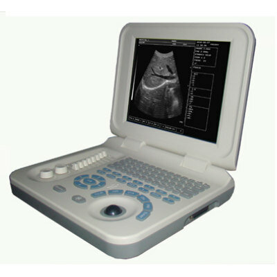
Laptop Ultrasound Scanner
PL-3018

Silver Member
Mobile X-Ray Barrier
Lead Acrylic Mobile X-Ray Barriers