MRI Cardiac Stress Test Could Help Identify CAD Severity
By MedImaging International staff writers
Posted on 20 Feb 2019
Researchers at Northwestern University (NU; Chicago, IL, USA), Heart Imaging Technologies (Durham, NC, USA), Duke University (Durham NC, USA), and other institutions conducted a study involving 9,151 patients (median age 63 years, 55% men) undergoing clinical evaluation of myocardial ischemia, in order to determine if stress CMR is associated with patient mortality. An automated process collected data from finalized clinical reports, aggregated the data, and assessed all-cause patient mortality via the US Social Security Death Index.Posted on 20 Feb 2019
The results revealed that 4,408 patients had a normal stress CMR examination, 4,743 had an abnormal examination, and 1,517 died during a median follow-up time of five years. Analysis revealed that for low-risk patients without prior history of heart disease, those with an abnormal CMR scan were 3.4 times more likely to die compared to patients with a normal CMR scan. The strong association between an abnormal stress CMR and mortality held even after adjusting for patient age, sex, and cardiac risk factors. The study was published on February 8, 2019, in JAMA Cardiology.
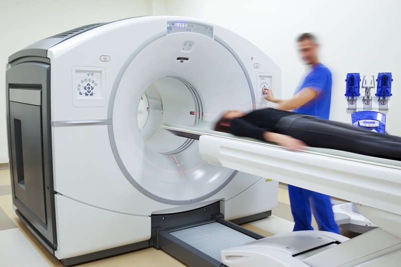
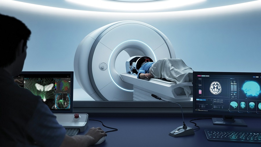
 could serve as a non-invasive, non-toxic alternative for identifying the extent of coronary artery disease (CAD) (Photo courtesy of Duke Health).")
Image: A new study suggests that cardiac magnetic resonance (CMR) could serve as a non-invasive, non-toxic alternative for identifying the extent of coronary artery disease (CAD) (Photo courtesy of Duke Health).
“CMR works as well or better than other exams at identifying heart wall motion, cell death, and the presence of low blood flow. There are a number of reasons for the limited use of stress CMR, including availability of good quality laboratories, exclusion of patients who cannot undergo magnetization, and a lack of data on patient outcomes,” said senior author Robert Judd, PhD, of Duke University. “With the findings from this study suggesting that stress CMR is effective in predicting mortality, we provide a strong basis for a head-to-head study between stress CMR and other modalities.”
Stress CMR perfusion is increasingly being used to test for inducible myocardial ischemia and has been well validated against other imaging modalities such as invasive angiography or fractional flow reserve (FFR). The majority of scans are performed using a stress/rest protocol using adenosine as the stressor, which acts to induce ischemia in the myocardium. Some centers use inotrope dobutamine to stress the heart, and the images are interpreted in a similar fashion to dobutamine stress echocardiogram.
Related Links:
Northwestern University
Heart Imaging Technologies
Duke University

Gold Member
Solid State Kv/Dose Multi-Sensor
AGMS-DM+

New
Ultrasound System
P20 Elite

Ultrasound Doppler System
Doppler BT-200

Ultrasound System
Acclarix AX9
.jpg)