MRI Effectively Monitors Liver Fat in Obese Patients
By MedImaging International staff writers
Posted on 02 Jan 2019
Measuring liver fat using magnetic resonance imaging (MRI) is a safe, noninvasive way to monitor the effects of bariatric surgery, according to a new study.Posted on 02 Jan 2019
Researchers at the University of Wisconsin (WISC; Madison, USA), the University of California, San Diego (UCSD, USA), and Virginia Commonwealth University (Richmond, USA) conducted a study involving 126 obese patients evaluated for bariatric surgery with a preoperative very low calorie diet (VLCD). All participants underwent chemical shift-encoded (CSE) MRI measurement of liver proton density fat fraction (PDFF) before VLCD, after VLCD, and 1, 3, and 6–10 months following surgery. Measures were rates of change of PDFF and body anthropometrics, including body mass index (BMI), weight, and waist circumference (WC).
.")
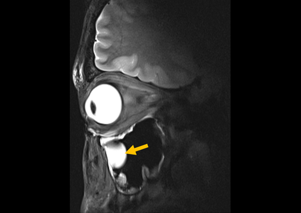
Image: PDFF maps in a 55-year-old woman who underwent gastric bypass demonstrate progressive reduction in liver fat (Photo courtesy of RSNA).
Of the initial 126 patients, 50 (mean age, 51 years; age range, 27–70 years), including 43 women and seven men completed the study. By six to 10 months after surgery, mean PDFF in the study group decreased from 18% to about five percent, and mean BMI decreased from 45 to 34.5 kg/m2. Mean estimated time to PDFF normalization was approximately five months. Initial PDFF was the only strong predictor for change in PDFF and time to PDFF normalization; no body anthropometric correlated with either outcome. The study was published on December 18, 2018, in Radiology.
“There is this assumption that when you lose weight you also reduce liver fat, but the relationship was very hard to measure prior to having a good tool like CSE-MRI, which allows us to represent the measurement of liver fat as a percentage," said study coauthor B. Dustin Pooler, MD, of WISC. “Each patient can get an assessment of fat throughout the liver that is easy for them to understand. The numbers also allowed us to perform comparisons with liver fat measurements from surgical and biopsy specimens.”
Bariatric surgical procedures such as gastric bypass or sleeve gastrectomy have proven to be effective weight loss interventions in patients with obesity. However, not much is known about the relationship between overall weight loss achieved by these treatments and decreases in liver fat content, as it is difficult to measure noninvasively. PDFF measurements could also help in the selection of patients for bariatric surgery because of the strong correlation between liver fat reductions and pre-treatment liver fat content.
Related Links:
University of Wisconsin
University of California, San Diego
Virginia Commonwealth University

Diagnostic Ultrasound System
DC-80A

Pocket Fetal Doppler
CONTEC10C/CL

High-Precision QA Tool
DEXA Phantom

Portable X-ray Unit
AJEX140H