Adaptable Contrast Media Improves MRI Diagnostics
By MedImaging International staff writers
Posted on 12 Dec 2018
An adaptable protein structure that absorbs dissolved xenon in a self-regulating way allows for higher quality magnetic resonance imaging (MRI) scans with less contrast medium, claims a new study.Posted on 12 Dec 2018
Developed by researchers at Leibniz-Forschungsinstitut für Molekulare Pharmakologie (FMP; Berlin, Germany) and the California Institute of Technology (Caltech; Pasadena, USA), the new contrast media is based on nanometric hollow protein structures produced by bacteria in order to regulate flotation depth, similar to a miniaturized swim bladder in fish. These so-called "gas vesicles" have a porous wall structure through which xenon gas can flow in and out, allowing them to elastically adjust their influence on the measured xenon.
.")
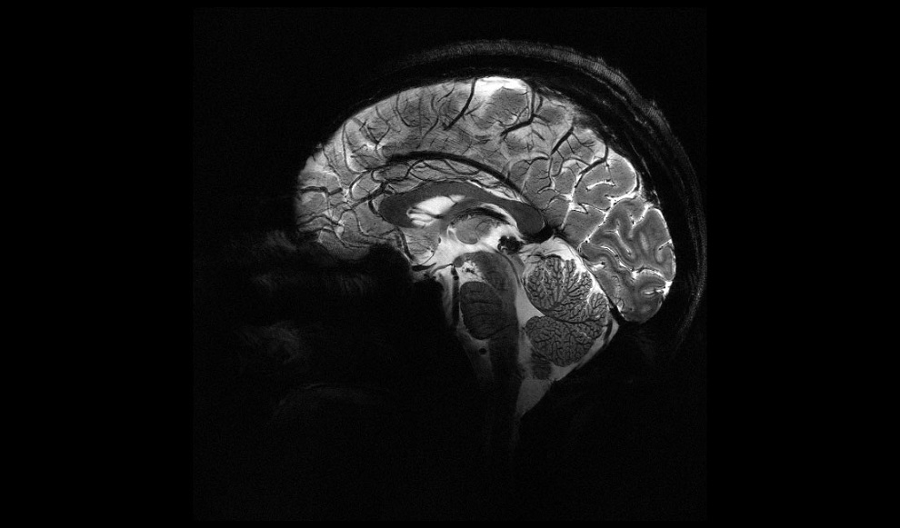
Image: Xenon filled vesicles provide higher contrast MRI scans than conventional contrast agents (Photo courtesy of Barth van Rossum / FMP).
Unlike conventional contrast media, the gas vesicles always absorb a fixed portion of the xenon present in the environment. This characteristic can be taken advantage of in MRI diagnostics, because more xenon gas must be used in order to obtain better images. While the concentration of a conventional contrast medium would need to be adjusted in order to achieve a change in signal for all xenon atoms, the gas vesicles automatically fill up with more xenon when this is offered.
As much more xenon is absorbed into the vesicles than with conventional contrast media, the image contrast is many times higher than the background noise, and image quality is significantly improved, allowing identification of disease markers that occur in relatively low concentrations. In animal studies, the researchers were able to produce MRI images with particle concentrations one million times lower than those of the contrast media currently employed. The study was published in the November 2018 issue of ACS Nano.
“We need new, improved methods, in which as little contrast medium as possible influences as much of the signal-transmitting substance as possible, typically water,” said senior author Leif Schroeder, PhD, director of the molecular imaging group at FMP. “They act like a kind of balloon, to which an external pump is attached. If the balloon is inflated by xenon atoms flowing into the gas vesicle its size does not change, but the pressure does increase, similar to a bicycle tire tube.”
Hyperpolarization of the nuclei of noble gases (usually using lasers) aligns them so that they become visible on an MRI scan. To detect specific cellular disease markers, they need to be bound to them for a short time. Gases used for medical imaging purposes include helium (He), argon (Ar), krypton (Kr), and xenon (Xe). The hyperpolarized spin state exists at very low spin temperatures, which leads to high magnetization of the spin ensemble, resulting in very high nuclear magnetic resonance signal intensity, which returns to thermal equilibrium by depolarization.
Related Links:
Leibniz-Forschungsinstitut für Molekulare Pharmakologie
California Institute of Technology

Gold Member
Solid State Kv/Dose Multi-Sensor
AGMS-DM+

New
Digital Radiography Generator
meX+20BT lite

New
Ceiling-Mounted Digital Radiography System
Radiography 5000 C
Portable X-Ray Unit
AJEX240H