Advanced Imaging Measures Iron Levels in Brain
By MedImaging International staff writers
Posted on 10 Dec 2018
A new study suggests magnetoencephalography (MEG) can be used to measure levels of magnetite in the human brain, which may help detect neurodegenerative disorders such as Alzheimer's disease (AD).Posted on 10 Dec 2018
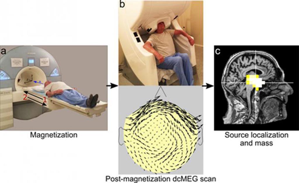
Researchers at Massachusetts General Hospital (MGH; Boston, USA) and the Massachusetts Institute of Technology (MIT; Cambridge, MA, USA) enrolled 11 male participants (19-89 years of age) who underwent an initial baseline direct current (dc) MEG scan before magnetic resonance imaging (MRI) scanning, both to acquire an image and to magnetize any magnetite particles within their brains. A second dcMEG scan was taken several minutes later to identify changes in the magnetic field, reflecting the size and shape of magnetite particles.

Image: Combining dcMEG data with an MRI shows magnetite location in the brain (Photo courtesy Sheraz Kahn/ MGH).
Subsequent alignment of the MEG and MRI images allowed precise localization of the magnetic signals. The results revealed greater accumulation of magnetite in the brains of the oldest volunteers, primarily in and around the hippocampus, replicating post-mortem studies. The rate at which the magnetic signal dissipated--a measure of particle size--was calculated by subsequent dcMEG scans taken from hours to several days later. The study was published on November 20, 2018, in Human Brain Mapping.
“The ability to measure and localize magnetite in the living brain will allow new studies of its role in both the normal brain and in neurodegenerative disease,” said study co-author David Cohen, PhD, of the MGH Martinos Center for Biomedical Imaging Center and MIT. “Studies could investigate whether the amount of magnetite in the hippocampal region could predict the development of Alzheimer's disease and whether treatments that influence magnetite could alter disease progression.”
“Our findings allow in‐vivo measurement of magnetite in the human brain, and possibly open the door for new studies of neurodegenerative diseases of the brain,” concluded lead author Sheraz Khan, PhD, of the MGH Martinos Center for Biomedical Imaging Center. “While this new tool is now ready to be applied in studies of patients with neurodegenerative diseases, several improvements, such as a new magnet specifically built for this purpose, will be required to produce the precise measurements required for accurate diagnosis.”
Magnetite (Fe3O4) particles in the human brain, first reported in 1992, draw strong interest due to their redox activity, strong magnetic behavior, particle surface charge, and Fenton‐like chemistry, which suggest they serve a physiological purpose in the normal brain. It has also been shown that magnetite is strongly associated with several degenerative diseases of the brain, especially AD, in which magnetite nanoparticles were found to be associated with tangles and plaques.
Related Links:
Massachusetts General Hospital
Massachusetts Institute of Technology


