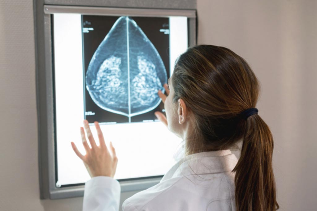
Automated System Identifies Dense Breast Tissue
By MedImaging International staff writers
Posted on 29 Oct 2018
A deep-learning (DL) automated model can assess dense breast tissue in mammograms as reliably as expert radiologists, claims a new study.Posted on 29 Oct 2018
Developed by researchers at the Massachusetts Institute of Technology (MIT, Cambridge, MA, USA) and Harvard Medical School (HMS; Boston, MA, USA), the DL model is based on a deep convolutional neural network (CNN) trained to assess breast imaging reporting and data system (BI-RADS) breast density--i.e. fatty, scattered, heterogeneous, and dense--based on the expert interpretation of 41,479 digital screening mammograms obtained in 27,684 women from January 2009 to May 2011. The algorithm was then tested on a held-out test set of 8,677 mammograms in 5,741 women.

Image: An artificial intelligence algorithm can detect dense breast tissue (Photo courtesy of MIT).
In addition, five radiologists performed a reader study on 500 mammograms randomly selected from the test set. Finally, the algorithm was implemented in routine clinical practice, where eight radiologists reviewed 10,763 consecutive mammograms assessed with the model. Agreement on BI-RADS category was compared for three sets of readings: radiologists in the test set, radiologists working in consensus in the reader study set, and radiologists in the clinical implementation set. The readings were compared across 5,000 bootstrap samples to assess significance.
The results revealed that the DL model showed good agreement with radiologists in the test set, and with radiologists in consensus in the reader study set. In addition, there was very good agreement with the radiologists in the clinical implementation set; for binary categorization of dense or non-dense breasts, 10,149 of 10,763 (94%) of DL assessments were accepted by the interpreting radiologist. On all four BI-RADS categories, the DL algorithm matched radiologists' assessments at 90%. The study was published on October 16, 2018, in Radiology.
“Breast density is an independent risk factor that drives how we communicate with women about their cancer risk. Our motivation was to create an accurate and consistent tool that can be shared and used across health care systems,” said study author PhD student Adam Yala, of the MIT Computer Science and Artificial Intelligence Laboratory (CSAIL). “When radiologists pull up a scan at their workstations, they'll see the model's assigned rating, which they then accept or reject. It takes less than a second per image ... [and it can be] easily and cheaply scaled throughout hospitals.”
It is estimated that more than 40% of women have dense breast tissue, which alone increases the risk of breast cancer. Moreover, dense tissue can mask cancers on the mammogram, making screening more difficult.
Related Links:
Massachusetts Institute of Technology
Harvard Medical School


