Virtual Heart Technology Guides Cardiac Ablation Procedures
By MedImaging International staff writers
Posted on 04 Oct 2018
A new study describes how personalized three-dimensional (3D) simulations can pinpoint infarct-related ventricular tachycardia (VT) ablation sites.Posted on 04 Oct 2018
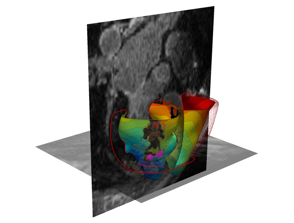
Developed by researchers at Johns Hopkins University (JHU; Baltimore, MD, USA), Simula Research Laboratory (Simula; Fornebu, Norway), the University of Utah (Salt Lake City, USA), and other institutions, the 3D personalized computational models are based on contrast-enhanced clinical magnetic resonance imaging (MRI) cardiac scans, with each virtual tissue cell generating electrical signals calculated using mathematical equations to represent which heart cells are healthy, and which are semiviable due to proximity to the infarction scar.

Image: A 3D simulated virtual heart (Photo courtesy of JHU).
By stimulating the patient’s virtual heart, the computer program can determine if the heart develops an arrhythmia, and the location of the tissue perpetuating it. The 3D model can then simulate an ablation to that area. The procedure can be repeated over and over again to find multiple ablation locations for the actual patient. For the study, the researchers first created personalized heart models of 21 people who underwent successful cardiac ablation procedures for infarct-related VT at JHU Hospital between 2006 and 2017. The 3D modeling correctly identified and predicted ablation sites.
In five of the patients, the amount of ablated tissue identified by the 3D model was smaller overall--in some cases, more than 10 times smaller--than the actual area destroyed during the procedures. The researchers then tested the 3D simulation to guide actual cardiac ablation treatments for another five patients. Most patients have remained free of VT throughout the follow-up period. In two patients, the virtual heart approach predicted that tachycardias would not be inducible, which was confirmed during the clinical procedure, so cardiac ablation was not performed. The study was published on September 3, 2018, in Nature Biomedical Engineering.
“Our new study results suggest we can remove a lot of the guesswork, standardize treatment, and decrease the variability in outcomes, so that patients remain free of arrhythmia in the long term,” said senior author Professor Natalia Trayanova, PhD, of the JHU department of biomedical engineering. “The approach could improve infarct-related VT ablation guidance, where accurate identification of patient-specific optimal targets could be achieved on a personalized virtual heart before the clinical procedure.”
In VT, the electrical signals in the heart’s lower chambers misfire, crippling the relaxation and refilling process and producing rapid arrhythmias. Numerous drugs are available to treat and manage infarct-related VT, but side effects and limitations of the drugs have increased focus on other interventions, especially cardiac ablation, which is successful anywhere between 50 and 88 percent of the time, but the outcomes are difficult to predict.
Related Links:
Johns Hopkins University
Simula Research Laboratory
University of Utah


