Dedicated System Scans Heart in One Heartbeat
By MedImaging International staff writers
Posted on 18 Sep 2018
A dedicated cardiovascular computerized tomography (CT) system creates a three-dimensional (3D) scan of the entire heart in one rotation.Posted on 18 Sep 2018
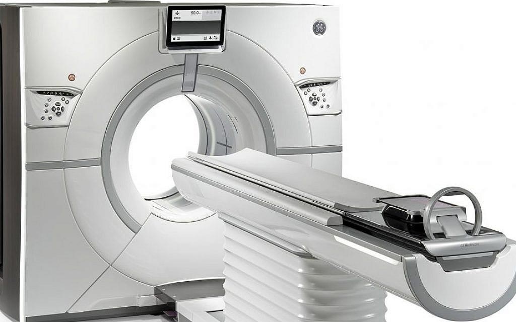
The GE Healthcare (GE, Little Chalfont, United Kingdom) Cardiographe is a proprietary CT scanner optimized for cardiovascular imaging by eliminating aspects required for whole-body imaging, but irrelevant for cardiac scanning, thus optimizing its operation for a range of cardiac imaging parameters. The compact, high performance system provides high resolution, motion-free, artifact-free, volumetric images of the whole heart without stitching, using a gantry rotation speed of 0.24 seconds, enabling very fast scans that freeze cardiac motion.

Image: A dedicated cardiovascular CT system targets the heart (Photo courtesy of Arineta).
Several innovative proprietary technologies are combined into the system to provide optimal cardiac imaging. The most important is focused field of view (FFW), which uses full beam intensity in the field of interest, and highly attenuated beam in the periphery. Two detector arrays are used, a high-resolution array in the focused field of view, and peripheral detector arrays optimized for low intensity imaging at the wings. Special algorithms are used to reconstruct the images in the center of the field of view, with GE SnapShot Freeze intelligent motion correction software integrated to suspend coronary motion.
Proprietary Twin Beam technology is also used, which involves two overlapping X-ray beams irradiating the scanned volume and detected both by a single detector. Ultra-fast beam switching technology and unique reconstruction algorithms optimize image quality. Finally, the system uses ultra short geometry (USG), wherein the twin x-ray source is substantially closer to the patient than in a conventional CT design. USG presents several advantages, including lower doses (2 mSv or below), faster rotation, and improved temporal resolution.
The Cardiographe was developed by Arineta (Ceserea, Israel), who partnered with GE Healthcare to commercialize the scanner. GE incorporated into the system features from its Revolution CT system, including advances for dose reduction, iterative reconstruction, metal artifact reduction and others. The Cardiographe was built to be very compact so it can fit into a physician’s office, or utilize a smaller room than a standard 64-slice scanner. The scanner is also optimized for other cardiovascular studies such as the aorta, the carotid, and renal arteries.
“It is more worthwhile for hospitals to buy one CT device at an average price for all the non-cardiac indications and our system for cardiac scans,” said Ehud Dafni, MD, CEO of Arineta. “That way, it can scan twice as many patients as it could by buying the expensive CT designed for all the systems, and which also has improved cardiac scan capabilities.”


