Functional Imaging System Identifies Neurological Disruptions
By MedImaging International staff writers
Posted on 04 Sep 2018
A sophisticated magnetoencephalography (MEG) system aids pre-surgical localization of epilepsy foci and mapping of the eloquent cortex, including motor functions, hearing, and vision.Posted on 04 Sep 2018
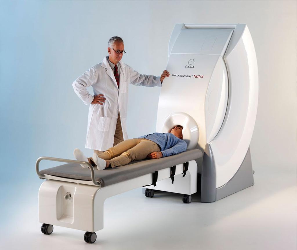
The Megin (Helsinki, Finland) Triux neo is a completely non-invasive diagnostic device designed to directly measure magnetic activity generated by neurons in the brain for diagnosis and assessment of complex neurological disruptions, with the ability to detect and localize neural events with millimeter accuracy and millisecond resolution. The system is intended for diagnosis of patients across a wide spectrum, including epilepsy, brain tumors, traumatic brain injury, post-traumatic stress disorder, and autism.

Image: A magnetoencephalography (MEG) system helps map neurological features (Photo courtesy of Megin).
The core technology is based on the ability to detect very weak magnetic fields--less than one-billionth the strength of the Earth’s magnetic field--in the brain using an array of superconducting quantum interference device (SQUID) array mounted in a close-fitting helmet cooled with liquid helium. Triux has 306 individual channels, making it effective for pre-surgical localization of epileptic foci, functional mapping of sensory, cortical and autonomic responses, revealing biomarkers for disease states or treatment responses, and research purposes.
Related Links:
Megin


