MRI Reveals Early Impact of Hypertension on Dementia Risk
By MedImaging International staff writers
Posted on 11 Jul 2018
A new study shows how magnetic resonance imaging (MRI) can reveal previously undetectable white matter (WM) brain damage induced by hypertension, a major risk factor for dementia.Posted on 11 Jul 2018
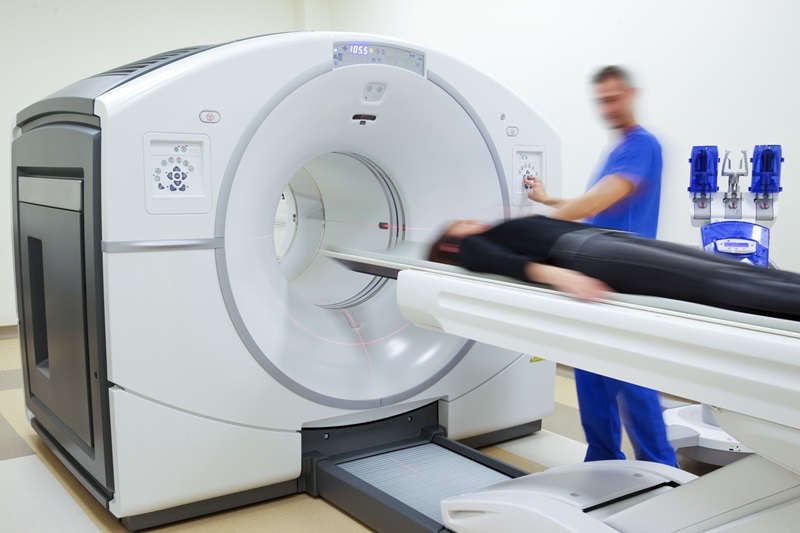
Researchers at IRCCS Neuromed (Pozzilli, Italy) and Sapienza University (Rome, Italy; www.uniroma1.it) conducted a study involving 42 outpatients (40-65 years of age), of whom 23 were hypertensive and 19 normotensive. The researchers first measured the cognitive performance of study participants with no signs of dementia or structural brain changes (as determined by a neuroradiologist), based on conventional MRI on T1 and AX DP/T2 scans. They then repeated imaging using WM fiber-tracking technology on a GE Healthcare (GE; Little Chalfont, United Kingdom; www.gehealthcare.com) 3T Signa Horizon scanner.
 and with (right) hypertension. The subject with hypertension had several deep and periventricular white matter lesions (arrows) (Photo courtesy of ResearchGate).")
Image: Cerebral MRIs of two women 67 years of age without (left) and with (right) hypertension. The subject with hypertension had several deep and periventricular white matter lesions (arrows) (Photo courtesy of ResearchGate).
In addition, a blinded, certified psychologist tested cognitive function and looked for any impairment of activities of daily living at baseline. Participants then underwent Montreal Cognitive Assessment (MoCA) neuropsychological testing. When specific MoCA cognitive subdomains were analyzed, the hypertensive group displayed significantly impaired memory, executive function, attention, and language domains, and scored worse on verbal paired-associate learning. Hypertension also appeared to impair executive function, as measured on the Stroop test.
When comparing WM finding between those with and without hypertension, specific signatures for dementia risk among hypertensive patients involved deterioration of projection fibers of the right anterior thalamic radiation (ATR), association fibers of the right superior longitudinal fasciculus (SLF), and callosum fibers of the forceps minor (FMI). The researchers concluded that WM tracking could identify patients in initial stages of brain damage that could benefit from therapies aimed at limiting the transition to dementia and neurodegeneration. The study was published on June 12, 2018, in Cardiovascular Research.
“All hypertensive participants were taking antihypertensive therapy, suggesting that an efficient anti-hypertensive therapy is not sufficient to exclude the onset of vascular dementia or protect the brain from white matter microstructural damage,” said lead author graduate student Lorenzo Carnevale, MSc, of NeuroMed. “Our results point out a big issue in current cardiovascular diseases treatment: We can't ignore the brain. Current therapies are not tailored to ensure at the same time cardiovascular protection and cognitive function protection.”
The ATR is a projection fiber that connects the thalamus's anterior nucleus to the anterior cingulate gyrus and frontal cortex, and ATR damage is associated with impaired memory. The SLF connects the cerebrum parietal and occipital and temporal lobes with the ipsilateral frontal cortices; it facilitates the formation of a bidirectional neural network necessary for cognitive functions, such as attention, memory, emotions, and language. The FMI connects the frontal lobes' lateral and medial surfaces, crosses the midline via the genu of the corpus callosum, and has been linked to processing speed tasks.
Related Links:
IRCCS Neuromed

Gold Member
Solid State Kv/Dose Multi-Sensor
AGMS-DM+

Color Doppler Ultrasound System
DRE Crystal 4PX

Silver Member
Mobile X-Ray Barrier
Lead Acrylic Mobile X-Ray Barriers

New
X-Ray QA Meter
Piranha CT
.jpg)