Laser-Sonic Scanner Images Entire Breast in Seconds
By MedImaging International staff writers
Posted on 25 Jun 2018
A new study describes how a photoacoustic computed tomography (PACT) scanning system can find breast tumors faster than mammography.Posted on 25 Jun 2018
Developed by researchers at Washington University School of Medicine (WUSTL; St. Louis, MO, USA), the California Institute of Technology (Caltech; Pasadena, USA), and other institutions, PACT involves shining a near-infrared (NIR) laser pulse that diffuses through the breast and is absorbed by oxygen-carrying hemoglobin molecules in the patient's red blood cells (RBCs), causing the molecules to vibrate ultrasonically. The vibrations travel through the tissue and are picked up by an array of 512 tiny ultrasonic sensors around the skin of the breast.

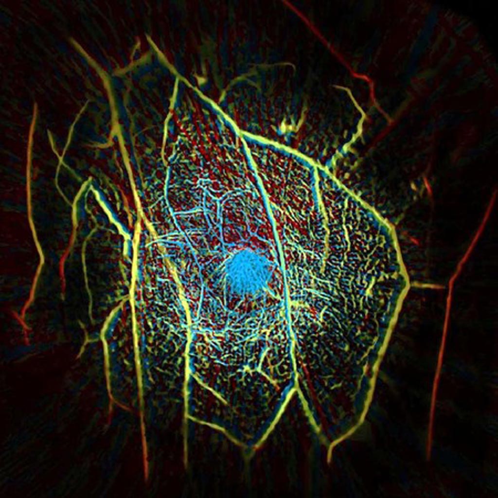
Image: The internal vascular structure of a human breast created using a PACT scanner (Photo courtesy of Caltech).
The data from the sensors are used to assemble an image of the breast's internal structures in a process that is similar to ultrasound imaging, though much more precise. Because the laser light is so strongly absorbed by hemoglobin, PACT can construct images that primarily show the blood vessels present in the tissue being scanned, providing a clear view of structures as small as a quarter of a millimeter at a depth of four centimeters.
Since the scan is quick, taking only 15 seconds, the patient can easily hold their breath while being scanned, and a clearer image can be developed. According to the researchers, the speed with which a PACT scan can be performed gives it advantages over other imaging techniques. For example, magnetic resonance imaging (MRI) scans can take 45 minutes, are expensive, and sometimes requires contrast agents to be injected into the patient's blood. The study was published on June 15, 2018, in Nature Communications.
“Mammograms cannot provide soft-tissue contrast with the level of detail in PACT images. This is the only single-breath-hold technology that gives us high-contrast, high-resolution, 3-D images of the entire breast,” said senior author professor of medical and electrical engineering Lihong Wang, PhD, of WUSTL. “Our goal is to build a dream machine for breast screening, diagnosis, monitoring, and prognosis without any harm to the patient. We want it to be fast, painless, safe, and inexpensive.”
PACT offers high spatial and temporal resolutions with sufficiently deep nonionizing optical penetration, since the principal optical absorber in the NIR region, hemoglobin, provides an endogenous contrast for imaging a high density of blood vessels. The concentration of blood vessels is often associated with angiogenesis, which plays an important role in tumor growth and metastasis.
Related Links:
Washington University School of Medicine
California Institute of Technology


