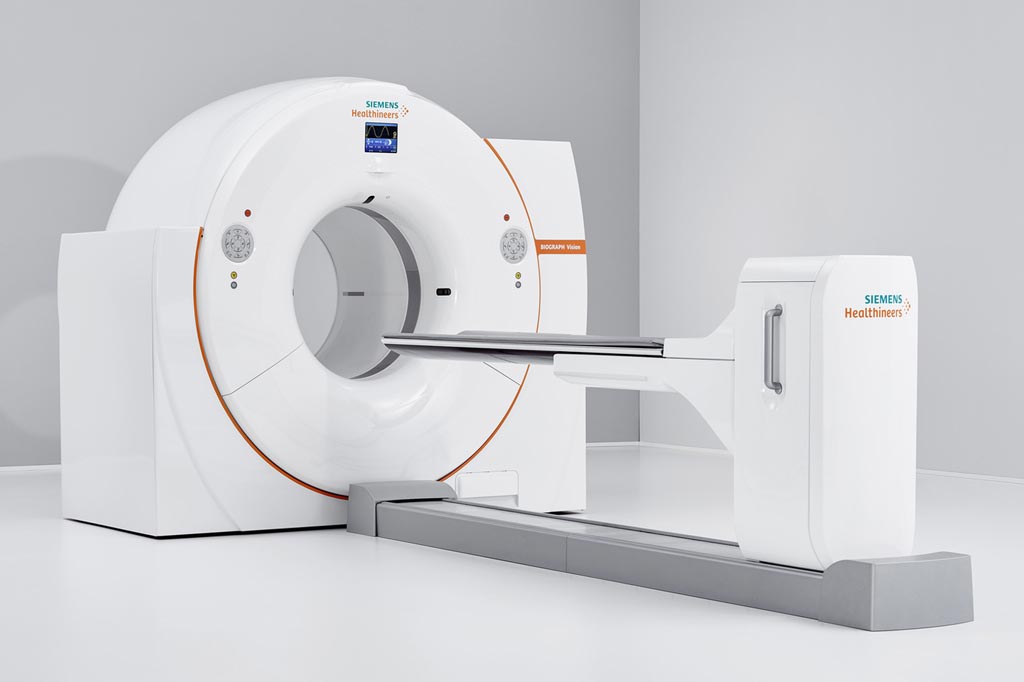
Innovative PET/CT System Improves Patient Comfort
By MedImaging International staff writers
Posted on 12 Jun 2018
A new positron emission tomography/computed tomography (PET/CT) system reduces scan time by a factor of 3.9, reducing patient radiation exposure and tracer cost.Posted on 12 Jun 2018
The Siemens Healthineers (Erlangen, Germany) Biograph Vision PET/CT system features Optiso ultra dynamic range (UDR) detectors, which uses silicon photomultipliers (SiPMs) rather than the standard photomultiplier tubes. Consequently, the detector’s lutetium oxyorthosilicate (LSO) crystal elements can be reduced from 4x4 mm to 3.2x3.2 mm, delivering higher spatial resolution. By utilizing the extremely small LSO crystals and covering 100% of the scintillator array area with SiPMs, the Biograph Vision can deliver extremely fast time-of-flight (ToF), with a temporal resolution of just 249 picoseconds, as well as a high effective sensitivity of 84 cps/kBq.

Image: The Biograph Vision PET/CT (Photo courtesy of Siemens Healthineers).
Features include QualityGuard, designed to self-calibrate the detector without an external radioactive source by tapping natural background radiation from the LSO detectors; FlowMotion Multiparametric Suite, a fully automated solution that delivers whole-body PET images of tracer uptake rate, metabolic glucose rate, and distribution volume, in addition to standard uptake volume (SUV) images; OncoFreeze, designed to provide images that are virtually free of respiratory motion; and CardioFreeze, which is designed to correct for respiratory and cardiac motion.
“The Biograph Vision represents a major leap in performance beyond any PET/CT system previously manufactured,” said Jim Williams, PhD, head of the Siemens Healthineers Molecular Imaging. “With this system, we extend the boundaries of PET imaging and help our customers explore a new frontier in precision medicine.”
PET is a nuclear medicine imaging technique that produces a three-dimensional (3D) image of functional processes in the body. The system detects pairs of gamma rays emitted indirectly by a positron-emitting radionuclide tracer. Tracer concentrations within the body are then constructed in 3D by computer analysis. In modern PET-CT scanners, 3D imaging is often accomplished with the aid of a CT X-ray scan performed on the patient during the same session, in the same machine.


