7T Scanner Provides Detailed Maps of Brain Activity
By MedImaging International staff writers
Posted on 08 May 2018
A new generation of highly sensitive 7 Tesla (7T) functional magnetic resonance imaging (fMRI) scanners can create precise maps of the brain before surgery so as to avoid damaging vital areas.Posted on 08 May 2018
A dynamic fMRI image correction process developed by researchers at the Medical University of Vienna (MedUni; Austria) for pre-surgery planning uses the 7T scanner multi-channel coils to create individual three-dimensional (3D) maps of the brain, with functional areas localized within the anatomy of the brain by using a dual-echo gradient echo reference scan and by calculating the phase offsets between the coils, which yields estimates with high signal-to-noise ratio.

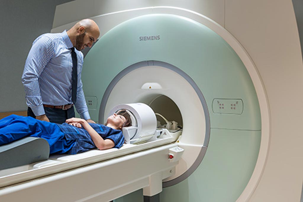
Image: In preparation for surgery, fMRI scanners can map cerebral blood flow (Photo courtesy of Siemens Healthcare).
Before the functional measurements start, the contribution of the 7T scanner to the measured signals is determined precisely. The resulting correction factor is then deducted from the fMRI images during the image computation. Neurologists can then decide whether surgery is useful, or even possible, and also determine which parts of the brain need to be spared at all costs. In a follow-up project, the researchers intend to develop methods to help determine the best possible location for deep brain stimulation (DBS) probes in patients suffering from Parkinson’s disease. A study describing the process was published in the March 2018 issue of Neuroimage.
“The higher magnetic field strength provides faster images of the brain functions at a higher resolution, but they are also more susceptible to distortions, which lead to imprecise functional imaging,” said lead author Simon Robinson, PhD, of the MedUni High Field MR Centre of Excellence. “In this way we are able to see, for instance, whether the language center has been displaced by a tumor. Unfortunately, the distortions of the magnetic field caused by bones, tissue and air are also stronger with 7T, which has implications for the mapping accuracy.”
Functional MRI measures brain activity by detecting changes associated with blood flow, based on the fact that cerebral blood flow and neuronal activation are coupled. Since the early 1990s, fMRI has come to dominate brain-mapping research because it does not require people to undergo shots or surgery, to ingest contrast agents, or be exposed to ionizing radiation. Using blood oxygen level dependent (BOLD) contrast, fMRI can localize activity to within millimeters, but only for a few seconds.
Related Links:
Medical University of Vienna


