Total-Body PET Creates New Patient Care Opportunities
By MedImaging International staff writers
Posted on 18 Jan 2018
A first-generation total-body positron emission tomography/computed tomography (PET/CT) scanner could revolutionize the understanding and treatment of disease, claims a new study.Posted on 18 Jan 2018
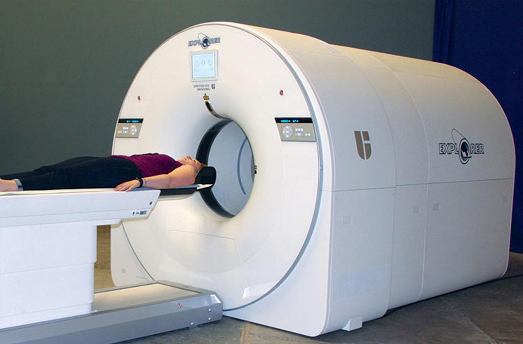
Under development at the University of California Davis (UCD, USA), in collaboration with United Imaging Healthcare (Shanghai, China), the prototype total-body PET/CT scanner, called EXPLORER, will offer the ability to detect focal pathologies throughout the whole body at once, including cancer, infection, and inflammation, and at considerably lower levels of disease activity than currently possible. It will also reduce the time taken to scan the whole body by at least a factor of 10, leading to scan times that could be less than one minute.

Image: The EXPLORER total-body PET/CT scanner (Photo courtesy of Martin Judenhofer /UCD).
By covering the entire body at once, sensitivity is increased by a factor of ~40 for total-body imaging, or a factor of ~4-5 for imaging a single organ such as the brain or heart. Significant improvements in timing resolution could lead to even further sensitivity gains. According to the researchers, the combination with total-body PET could produce overall sensitivity gains of more than two orders of magnitude, when compared to existing state-of-the-art systems. The study was published in the January 2018 issue of the Journal of Nuclear Medicine (JNM).
“One total-body PET scanner could take on the work load of three-to-four conventional PET scanners, and being able to receive imaging biomarkers from more distant distribution centers will minimize the need for costly in-house biomarker production,” said Terry Jones, DSc, clinical professor of diagnostic radiology at UCD. “As the impact of high-sensitivity, total-body PET scanning becomes apparent, this will provide a major stimulus to physicists, chemists, and engineers to develop lower-cost detectors for total-body surveillance.”
PET is a nuclear medicine imaging technique that produces a 3D image of functional processes in the body. The system detects pairs of gamma rays emitted indirectly by a positron-emitting radionuclide tracer. Tracer concentrations within the body are then constructed in 3D by computer analysis. In modern PET-CT scanners, 3D imaging is often accomplished with the aid of a CT X-ray scan performed on the patient during the same session, in the same machine.
Related Links:
University of California Davis
United Imaging Healthcare


