Neurofeedback Shows Promise as Tinnitus Treatment
By MedImaging International staff writers
Posted on 13 Dec 2017
Functional MRI (fMRI) demonstrates that neurofeedback training (NFT) has the potential to reduce the severity of tinnitus or even eliminate it, according to a new study.Posted on 13 Dec 2017
Researchers at Wright State University (Fairborn, OH, USA) conducted a clinical study involving 18 healthy volunteers with normal hearing in order to determine the potential efficacy of self-regulation of the primary auditory cortex to treat tinnitus via real-time fMRI neurofeedback; volunteers underwent five fMRI-NFT sessions, composed of an initial simple auditory fMRI followed by two runs of auditory cortex fMRI-NFT. fMRI results were recorded using single-shot echoplanar imaging, an MRI technique that is sensitive to blood oxygen levels, providing an indirect measure of brain activity.

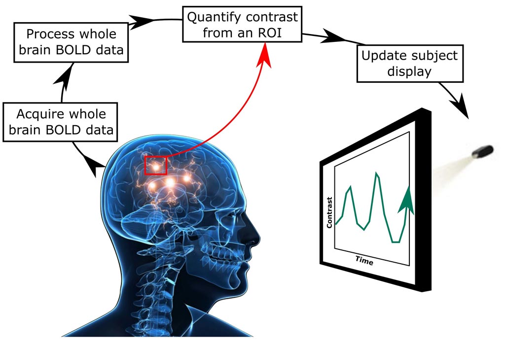
Image: The standard approach to fMRI neurofeedback (Photo courtesy of the Radiological Society of North America).
The simple auditory fMRI was taken in an MRI scanner, with the volunteers wearing noise-canceling earplugs. The run was comprised of six blocks containing a 20 second period of no auditory stimulation, followed by a 20 second period of white noise stimulation at 90 dB. Auditory cortex activity was then defined from a region using the activity during the preceding auditory run, and continuously updated during fMRI-NFT using a simple bar plot, accompanied by 90 dB white noise stimulation for the duration of the scan.
The participants then participated in the fMRI-NFT phase, receiving white noise through their earplugs while viewing activity in their primary auditory cortex as a bar on a screen. Each fMRI-NFT run contained eight blocks separated into a 30 second relax period followed by a 30 second lower period. Volunteers were instructed to watch the bar during the relax condition, and actively lower the bar by decreasing auditory cortex activity. Many participants focused on breathing, as it gave them a feeling of control, and diverted their attention away from sound. The study was presented at the annual meeting of the Radiological Society of North America (RSNA), held during November 2017 in Chicago (IL, USA).
“The idea is that in people with tinnitus there is an over-attention drawn to the auditory cortex, making it more active than in a healthy person,” said lead author and study presenter Matthew Sherwood, PhD, of the department of biomedical, industrial, and human factors engineering. “Our hope is that tinnitus sufferers could use neurofeedback to divert attention away from their tinnitus and possibly make it go away. Ultimately, we'd like take what we learned from MRI and develop a neurofeedback program that doesn't require MRI to use, such as an app or home-based therapy that could apply to tinnitus and other conditions.”
Tinnitus is the perception of sound within the human ear when no actual sound is present. It is not a disease, but a condition that can result from a wide range of underlying causes, including neurological damage, ear infections, oxidative stress, foreign objects in the ear, nasal allergies, wax build-up, and exposure to loud sounds. While it may be an accompaniment of sensorineural hearing loss or congenital hearing loss, or a side effect of certain medications, the most common cause is noise-induced hearing loss. Tinnitus is common, with about 20% of people between 55 and 65 years old report symptoms on a general health questionnaire, and 11.8% on more detailed tinnitus-specific questionnaires.
Related Links:
Wright State University


 Guided Devices.jpg)


