Photon Camera Sees through Human Body
By MedImaging International staff writers
Posted on 22 Sep 2017
A new imaging technique can overcome the limitations imposed by tissue scattering to optically determine the location of fiberoptic medical devices.Posted on 22 Sep 2017
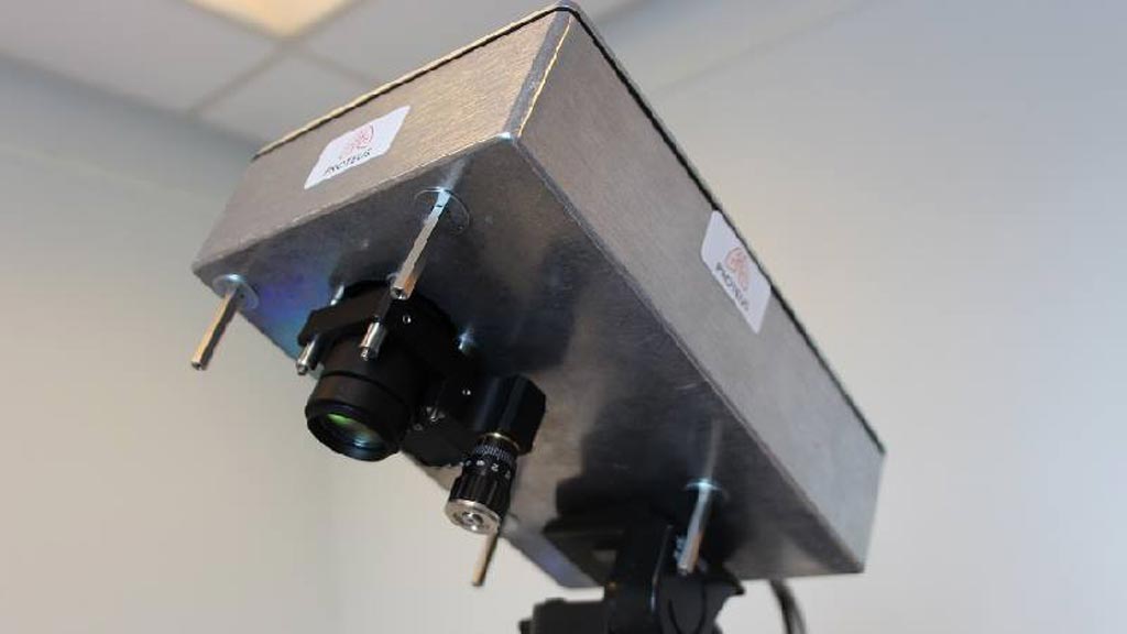
Developed by researchers at Heriot-Watt University (Edinburgh, United Kingdom) and the University of Edinburgh (United Kingdom), the camera is based on a time resolved detector array that integrates thousands of single photon detectors onto a single silicon chip, similar to that found in a digital camera. The array is used to identify ballistic and snake photons, which help determine the exact location of the distal end of the fiberoptic device that is emitting the light deep inside tissue.

Image: A novel camera can detect photons passing through the body (Photo courtesy of Heriot-Watt University).
The technology is so sensitive that it can detect the photons passing through the body's tissue. And by taking into account both the scattered light, the light that travels straight to the camera, and the elapsed time, the device is able to work out exactly where the endoscope is located in the body. Early tests show that the prototype device can track the location of a point light source through 20 centimeters of tissue under normal light conditions with centimeter resolution. The study was published in the September 2017 issue of Biomedical Optics Express.
“This is an enabling technology that allows us to see through the human body. It has immense potential for diverse applications such as the one described in this work,” said study co-author Professor Kev Dhaliwal, MD, PhD, of the University of Edinburgh. “The ability to see a device's location is crucial for many applications in healthcare, as we move forwards with minimally invasive approaches to treating disease.”
“My favorite element of this work was the ability to work with clinicians to understand a practical healthcare challenge, then tailor advanced technologies and principles that would not normally make it out of a physics lab to solve real problems,” said lead author Michael Tanner, MD, of Heriot-Watt University. “I hope we can continue this interdisciplinary approach to make a real difference in healthcare technology.”
Related Links:
Heriot-Watt University
University of Edinburgh


