Palette of Fluorescent Molecules Advance Biological Imaging
By MedImaging International staff writers
Posted on 15 Sep 2017
A new technique can create a colorful spectrum of fluorescent dyes by utilizing specific chemical molecules called rhodamines, claims a new study.Posted on 15 Sep 2017
The new dyes were developed by researchers at the Howard Hughes Medical Institute (HHMI; Ashburn, VA, USA), who previously developed the Janelia Fluor (JF) series of enhanced fluorescent dyes that are characterized by substantial increases in both brightness and photostability, achieved by incorporating four-member azetidine rings into classic fluorophore structures. They have now further refined and extended the strategy by incorporating 3-substituted azetidine groups, which allows rational tuning of the spectral and chemical properties of rhodamine dyes.

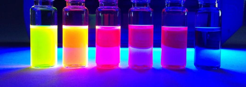
Image: Rhodamine dyes fluorescing under ultraviolet illumination (Photo courtesy of Jonathan Grimm/ HHMI).
The new strategy allowed them to establish principles for fine-tuning the properties of fluorophores and to develop a palette of new fluorescent and fluorogenic labels with excitation ranging from blue to the far-red. For the study, the researchers lit up cell nuclei, made larval fruit fly brains shine, and highlighted visual cortex neurons in mice that had tiny glass windows fitted into their skulls. And as the new dyes are synthesized in a single step with inexpensive ingredients, they are much cheaper than commercial alternatives. The study was published on September 4, 2017, in Nature Methods.
“By carefully placing just a few new atoms in the dye structure, the color and chemical properties of the dyes could also be fine-tuned, allowing many shades of green from a single scaffold; It's like going from the classic eight pack of crayons to the jumbo box of 64,” said senior author Luke Lavis, PhD, of the HHMI Janelia research campus. “Now we've figured out the rules, and we can make almost any color. Select the right atoms and chemists can engineer dyes with nearly any property they want.”
Until about 20 years ago, scientists relied on chemical fluorescent dyes to make biological molecules visible. But in 1994, a genetic trick was developed to tack green fluorescent protein (GFP), found in jellyfish, onto other cellular proteins, allowing researchers to trace protein movement without using expensive synthetic dyes. The discovery and development of GFP earned the Nobel Prize in chemistry for three scientists, including the late Professor Roger Tsien, PhD, a UCSD professor of pharmacology, chemistry, and biochemistry.
Related Links:
Howard Hughes Medical Institute


