Novel Targeted Tracer Helps Image Pending Aneurysms
By MedImaging International staff writers
Posted on 16 Aug 2017
A new single-photon emission computed tomography/computed tomography (SPECT/CT) imaging tracer may help assess abdominal aortic aneurysm (AAA) rupture risk, according to a new study.Posted on 16 Aug 2017
Developed at Yale University School of Medicine (New Haven, CT, USA) and the Veterans Affairs Connecticut Healthcare System (VACT; West Haven, USA), the new tracer, RYM1, is based on a water-soluble zwitterionic matrix metalloproteinase (MMP) inhibitor, which is coupled with 6-hydrazinonicotinamide and labeled with 99mTc. In studies in mice, the 99mTc-RYM1 tracer was radiochemically stable in blood for five hours, demonstrating rapid renal clearance and lower blood levels in vivo compared with 99mTc-RP805.

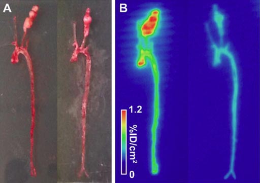
Image: A novel imaging tracer can help detect risk of abdominal aortic aneurysm (Photo courtesy of Yale University).
In radiographic studies, specific binding of 99mTc-RYM1 to aneurysm and its specificity were shown in a carotid aneurysm, with in-vivo SPECT/CT images showed higher uptake of the tracer in AAA than non-dilated aortas. The researchers also demonstrated that uptake of 99mTc-RYM1 correlated with aortic MMP activity and macrophage marker CD68 expression, as assessed by zymography and reverse-transcription polymerase chain reaction (PCR). The study was published in the August 2017 issue of the Journal of Nuclear Medicine.
“There is no effective medical therapy for AAA, and current guidelines recommend invasive repair of large AAA. However, the morbidity and mortality remain high, so better tools for AAA risk stratification are needed,” said senior author Mehran Sadeghi, MD, of the Yale Cardiovascular Research Center (CRC). “Further development of RYM1-based imaging could expand the applications of molecular imaging and nuclear medicine, and improve patient management in a wide range of diseases.”
MMPs are a group of enzymes that in concert are responsible for the degradation of most extracellular matrix (ECM) proteins during organogenesis, growth, and normal tissue turnover. While expression and activity of MMPs in adult tissues is normally quite low, it increases significantly during some pathological conditions that lead to unwanted tissue destruction, such as inflammatory diseases, tumor growth, and metastasis. MMPs play a key role in AAA development.
Related Links:
Yale University School of Medicine
Veterans Affairs Connecticut Healthcare System


