New OCT Technique Images Cellular Structure of Eye
By MedImaging International staff writers
Posted on 16 Aug 2017
A new study describes how linear optical coherence tomography (OCT) allows clinicians to resolve individual photoreceptors, capillary blood vessels, and nerve fibers in the same image.Posted on 16 Aug 2017
Developed at the Medical University of Vienna (MedUni; Austria) Line Field OCT uses noniterative digital aberration correction (DAC) to achieve aberration-free cellular-level resolution in OCT images of the human retina in vivo. The system is based on a line-field spectral-domain OCT system with a high tomogram rate. The researchers also applied DAC on functional OCT angiography data in order to improve lateral resolution and compensate for defocus.

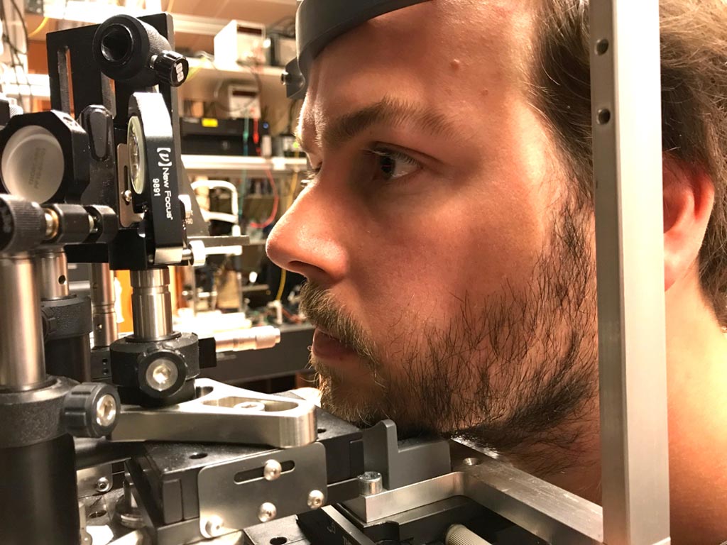
Image: A new OCT technique provides better 3D imaging of the eye (Photo courtesy of MedUni Vienna).
Functionally, the Line Field OCT is similar to a scanner, focusing a thin linear beam of light onto the internal structures of the eye. With speeds reaching up to 2.5 kHz, DAC can be applied not only to image human cone photoreceptors, but also to obtain an aberration- and defocus-corrected three-dimensional (3D) volume. DAC speed necessities were measured by examining the axial motion of the OCT system in 36 subjects, with the aim of appropriately quantifying motion analysis. The study was published in the August 2017 issue of Optica.
“Our new technique enables us to make digital corrections without the need for expensive hardware-based adaptive lenses. The linear illumination that is used allows very rapid frame rates, which are extremely important for these corrections,” said lead author Laurin Ginner, MSc, of the MedUni Center for Medical Physics and Biomedical Engineering. “This enables us to correct aberrations over the entire three-dimensional volume of the retina.”
OCT is based on low-coherence interferometry, typically employing near-infrared (NIR) light. The use of relatively long wavelength light allows it to penetrate into the scattering medium. Depending on the properties of the light source, OCT can achieve sub-micrometer resolution. OCT, being an echo imaging method, is similar to ultrasound imaging, but is limited to 1-2 mm below the surface in biological tissue, as at greater depths the proportion of light that escapes without scattering is too small to be detected.
Related Links:
Medical University of Vienna


