New Endoscope Targets Colon Tumors
By MedImaging International staff writers
Posted on 18 May 2017
An innovative endoscope will use breakthrough photonics technology to enable rapid, accurate diagnosis of bowel polyps and early colon cancer.Posted on 18 May 2017
The PhotonICs endoscope for improved in-vivo COLOn Cancer diagnosis and clinical decision support (PICCOLO), under development at Tyndall National Institute, Tecnalia, and other institutions, is a multimodal photonic endoscope with high sensitivity that will use both optical coherence tomography (OCT) and multi-photon tomography (MPT), combined with fluorescence technology, in order to facilitate real time computer aided diagnosis (CAD) of colon cancer via high-resolution structural and functional imaging.

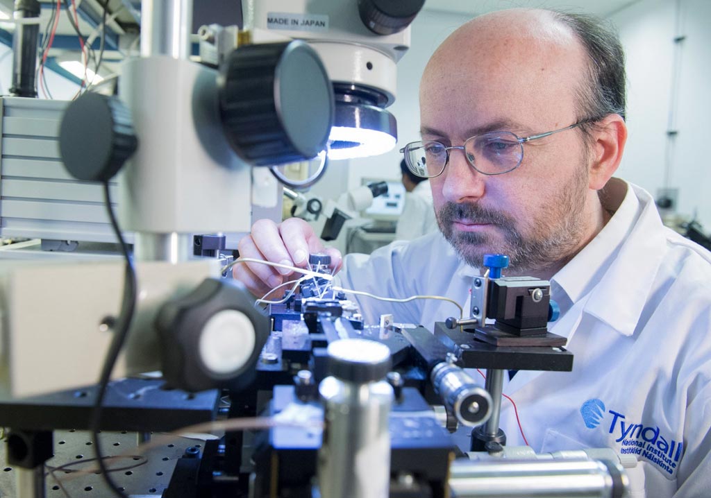
Image: The new endoscope will enable faster, more accurate colorectal cancer diagnosis (Photo courtesy of the Tyndall National Institute).
The PICCOLO will be able to detail changes occurring at the cellular level, comparable to those obtained using traditional histological techniques. According to the researchers, the endoscope will not only provide a new approach in colon cancer detection, but its image-based diagnosis methods could be applied to diseases in other organs of the body. The PICCOLO multidisciplinary development team, which includes clinicians, photonics components and technology providers, endoscope providers, and medical software developers hope to have refined the first prototype by the end of 2018, and target clinical trials to begin around 2020.
“When multiple polyps are detected in a patient, the current gold standard procedure is to remove all of them, followed by microscopic tissue analysis,” said computer vision researcher Artzai Picon, PhD, of Tecnalia. “Removal of hyperplastic polyps, which carry no malignant potential, and the subsequent costly histolopathological analysis might be avoided through the use of the PICCOLO endoscope probe, which could allow image-based diagnosis without the need for tissue biopsies.”


