Digital Tomosynthesis Reduces Re-Excision in Breast Surgeries
By MedImaging International staff writers
Posted on 20 Apr 2017
A new study shows that digital breast tomosynthesis (DBT) can reduce surgical positive margins by offering a better view of lesion margins than mammography alone.Posted on 20 Apr 2017
Researchers at the University of Turin conducted a study of 925 patients who had breast surgery between 2010 and 2015. Of these, 685 were breast-conserving surgeries, while 240 were mastectomies. Prior to surgery, 537 patients were staged with digital mammography and ultrasound, and 388 were staged with digital mammography, ultrasound, and DBT. In the first group, 348 women had lesions two cm or smaller, and 189 had lesions larger than two cm; in the second group, 207 women had lesions two cm or smaller, and 181 had lesions larger than two cm.

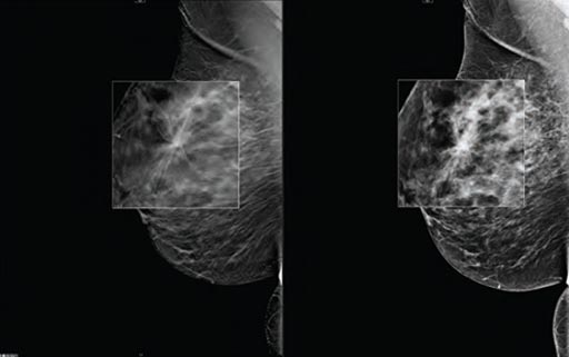
Image: A new study shows digital breast tomosynthesis reduces cancer surgery re-excision rates (Photo courtesy of Carestream Health).
Histology revealed that 7.9% of the patients had positive margins at histology. The re-excision rate for the first group was 10%, significantly higher than for the second group, who underwent DBT as well, which was 5%. Re-excision planning was also less extensive for patients in the second group, with one of 20 patients receiving a mastectomy, compared with 20 of 53 patients having mastectomies in the first group. The re-excision rate for lesions smaller than two cm was not statistically significant between the two groups, in contrast to larger lesions. The study was presented at ECR 2017, held during March 2017 in Vienna (Austria).
“Margin status is one of the most important predictors for local recurrence following breast cancer surgery, and accurate pre-operative staging helps to plan appropriate surgical treatment and reduce the consequences of re-excision,” said lead author and study presenter Alessia Milan, PhD. “These consequences can include emotional burden for the patient, a worse cosmetic outcome and higher costs. Our findings suggest that DBT has a role in improving conventional imaging for preoperative breast cancer surgical treatment staging.”
DBT mammograms use low dose x-rays to create a three-dimensional (3D) image of the breast, which can then be viewed the in narrow slices, similar to computerized tomography (CT) scans. While in conventional 2D mammography overlapping tissues can mask suspicious areas, 3D images eliminate the overlap, making abnormalities easier to recognize. It is estimated that 3D DBT will replace conventional mammography within ten years.


