Laser Microscopy Helps Examine Blood Vessels in Brain
By MedImaging International staff writers
Posted on 18 Jan 2017
An innovative technique to perfuse the brain vasculature helps quantify blood vessels using three dimensional (3D) image analysis procedures.Posted on 18 Jan 2017
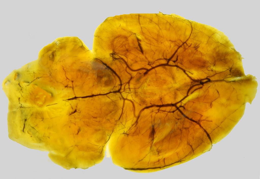
Developed by researchers at the University of Surrey and the Federal University of São Paulo, the technique involves dissolving China Ink with gelatin to create a solution that makes blood vessels more visible when viewed with a confocal microscope, enabling clinicians and pathologists to make an accurate reading of the number, length, and surface area of the brain vasculature. They can also create 3D images, which can help identify changes in their shape and size, key indicators of a number of circulation-related diseases of the brain.

Image: A mouse brain imbued with China Ink to enhance vasculature (Photo courtesy of the University of Surrey).
The innovative technique, a modification of the so-called Spalteholz method, will also facilitate a greater understanding of how exercise affects the brain by examining the circulatory effects of increased or decreased heart rate and arterial pressure on the brain. It will also help in investigating angiogenesis in cortical, hippocampal, and cerebellar tissue samples, including stereological estimations of total volume and length and of surface area of vessels. The study was published on January 5, 2017, in the Journal of Anatomy.
“Previously, we have been unable to fully sample and perform a quantification of the circulation of the brain in 3D as we simply could not see all vessels, due to their minute size and sometimes due to their irregular spatial distribution,” said study co-author Augusto Coppi, MD, of the University of Surrey. “This new technique will allow us to sample, image, and count blood vessels in 3D, giving us a greater mechanistic comprehension of how the circulation of the brain works, and how brain diseases such as dementia and stroke affect this organ.”
Confocal laser-scanning microscopy is a useful tool for visualizing neurons and glia in transparent preparations of brain tissue from laboratory animals. Currently, imaging capillaries and venules in transparent brain tissues requires the use of fluorescent proteins.


