Radiation-Free Approach Images Molecules in Brain
By MedImaging International staff writers
Posted on 22 Dec 2016
A new study describes how protein-based sensors can cause blood vessels to dilate near their targets, allowing them to be detected by magnetic resonance imaging (MRI).Posted on 22 Dec 2016
Developed by researchers at the Massachusetts Institute of Technology (MIT, Cambridge, MA, USA), the probes are based on a modified calcitonin gene-related peptide (CGRP), which is active primarily during migraines or inflammation. The novel CGRP-related peptide is used to artificially activate vasculature dilation at nanomolar concentrations, thus enhancing an endogenous multimodal tissue contrast which can then be readily measured by optical and MRI scans in order to determine where the proteases were detected.

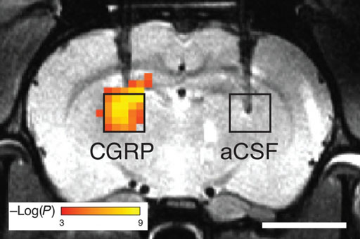
Image: CGRP-based sensors dilate blood vessels in the brain to make them visible on MRI (Photo courtesy of Alan Jasanoff / MIT).
In order to direct the CGRP-related peptide to their targets, they have been engineered so that they are trapped within a protein enclosure that keeps them from interacting with blood vessels. But when the peptides encounter proteases in the brain, the enclosures are disrupted, releasing CGRP, which causes nearby blood vessels to dilate. The researchers are now working on adapting the design to monitor neurotransmitters by modifying the enclosures surrounding the CGRP so that they can be removed by interaction with a specific neurotransmitter. The study was published on December 2, 2016, in Nature Communications.
“Currently the gold standard approach to imaging molecules in the brain is to tag them with radioactive probes. However, these probes offer low resolution and they can't easily be used to watch dynamic events,” said lead author professor of biological engineering Alan Jasanoff, PhD. “This is an idea that enables us to detect molecules that are in the brain at biologically low levels, and to do that with these imaging agents or contrast agents that can ultimately be used in humans.”
The vasculature is one of the most potent endogenous contrast sources available to imaging modalities. Vascular hemodynamic changes can be evoked by a variety of chemical species, many of which act at nanomolar concentrations. The changes in blood volume, flow, or oxygenation are robustly detectable by MRI, optical, and ultrasound-based noninvasive imaging, as well as by positron emission tomography (PET), single photon computed tomography, and X-ray imaging with intravascular tracers.
Related Links:
Massachusetts Institute of Technology


