Cardiac PET/CT Imaging Effective in Detecting Arterial Calcium
By Daniel Beris
Posted on 08 Dec 2016
A new study shows that cardiac positron emission tomography/computed tomography (PET/CT) imaging is effective in measuring total coronary artery calcification.Posted on 08 Dec 2016
Researchers at Intermountain Medical Center Heart Institute (Murray, UT, USA) conducted a study of 658 patients (mean age 67.2 years) who passed a stress test, and who went on to have a clinically indicated regadenoson rubidium-82 PET/CT scan and concurrent coronary artery calcification quantification. Calcium presence and density were measured through an Agatston score, which was defined as mild for a score of 0-10, moderate for a score of 11-299 as moderate, and severe as a score of 300-1,000 as severe. Scores higher than 1,000 were ruled as very severe.

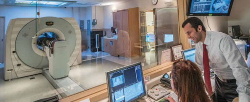
Image: Research shows cardiac PET/CT imaging is effective in detecting coronary calcium (Photo courtesy of Intermountain Medical Center Heart Institute).
The results showed a significant correlation between the amount of calcium and the occurrence of cardiac events; 3.88% of the patients with moderate calcification, 5.26% of the patients with severe calcification, and 7.14% of the patients with very severe calcification had a cardiac event within a year. In all, 16.28% of patients with coronary artery calcification suffered a cardiac event during the study period. The 33 patients (5%) with no or mild calcification had no cardiac events. The study was presented at the American Heart Association (AHA) annual meeting, held during November 2016 in New Orleans (LA, USA).
“People say 'I'm good -they gave me a stress test', but it doesn't tell the whole story. The story it tells is that on that day your engine - your heart - passed the test. Some of these people die within a year from a heart attack,” said lead author and study presenter Viet Le, MPAS. “Cardiac PET provides so much more for so much less radiation exposure, and with CT we can get anatomical imaging on a scanner that also gives us functional imaging.”
PET is a nuclear medicine imaging technique that produces a three-dimensional (3D) image of functional processes in the body. The system detects pairs of gamma rays emitted indirectly by a positron-emitting radionuclide tracer. Tracer concentrations within the body are then reconstructed in 3D by computer analysis. In modern PET-CT scanners, 3D imaging is often accomplished with the aid of a CT X-ray scan performed on the patient during the same session, in the same machine.
Related Links:
Intermountain Medical Center Heart Institute


