Novel Activity-Based Probes Detect Vascular Inflammation
By MedImaging International staff writers
Posted on 08 Nov 2016
Researchers have shown that a combined optical and Positron Emission Tomography (PET)/Computed Tomography (CT) probe can be used to detect early signs of atherosclerotic plaques.Posted on 08 Nov 2016
The researchers published the study in the October 2016, issue of the Journal of Nuclear Medicine. Atherosclerosis is mostly asymptomatic and can develop over many years. Symptoms only become apparent when more than 70% of a vessel is blocked resulting in a significant risk of a stroke or myocardial infractions.

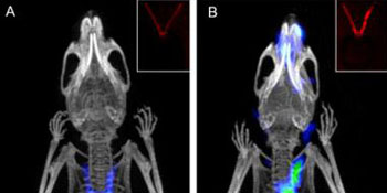
Image: The images demonstrate the use of activity-based probes using optical and PET/CT methods, to detect early signs of atherosclerotic plaques (Photo courtesy of Xiaowei Ma, Toshinobu Saito and Nimali Withana).
The researchers from the Stanford University Medical Center (SUMC; Stanford, CA, USA) used Activity-Based Probes (ABPs) and optical and PET/CT methods. The researchers were able to demonstrate that ABPs, which target cysteine cathepsins provide a fast and non-invasive technique to enable them to image the progression of atherosclerotic disease and the vulnerability of plaque.
One of the lead authors of the study, Matthew Bogyo, PhD, said, "This collaborative study with Zhen Cheng, PhD, and Michael McConnell, MD, provides evidence that these probes have potential benefits for non-invasive imaging of atherosclerotic plaque inflammation, potentially leading to the application of this probe in the clinic to help identify patients at high risk of developing premature atherosclerosis. What's novel about this is the fact that these probes provide accurate detection of lesions undergoing high levels of inflammatory activity and extracellular matrix remodeling. They not only enable early disease detection, they can provide real-time monitoring of therapeutic responses and clinical drug efficacy."
Related Links:
Stanford University Medical Center


