Nanoparticle Imaging Agents Reveal Microdamaged Bone Structure
By MedImaging International staff writers
Posted on 22 Sep 2016
A revolutionary bone-scanning technique produces extremely high-resolution three-dimensional (3D) images of bone, without exposing patients to X-ray radiation.Posted on 22 Sep 2016
Developed by researchers at Trinity College Dublin (Ireland), in collaboration with The Royal College of Surgeons in Ireland (RCSI; Dublin, Ireland), the technique is based on Europium-emitting surface-modified gold nanoparticles (AuNP) that serve as a contrast agent for fluoroscopy imaging of micro-damaged bone structure. The nanostructures can be utilized to generate 3D maps of the cracks formed within damaged bone, since the Europium nanoagents are attracted to calcium-rich surfaces.

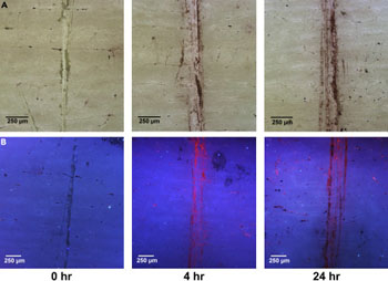
Image: Microdamaged bovine bone in polarized light (A), and after being immersed in an aqueous solution of AuNP (B) (Photo courtesy of Trinity College).
Using two-photon excitation fluorescence microscopy, the researchers were able to visualize and understand the manner in which the nanoagents bind to damaged bone, as well as demonstrate their selectivity toward exposed calcium at low concentrations. According to the researchers, the technique will have major implications for the health sector, as it can be used to diagnose bone strength and provide a detailed blueprint of the extent and precise positioning of any weakness or injury. The study was published on September 8, 2016, in Chem.
“We have demonstrated that we can achieve a 3D map of bone damage, showing the so-called microcracks, using non-invasive luminescence imaging,” said senior author professor of chemistry Thorri Gunnlaugsson, PhD. “The nanoagent we have developed allows us to visualize the nature and the extent of the damage in a manner that wasn't previously possible. This is a major step forward in our endeavor to develop targeted contrast agents for bone diagnostics for use in clinical applications.”
“Current X ray techniques can tell us about the quantity of bone present, but they do not give much information about bone quality,” added professor of anatomy Thomas Clive Lee, MD, PhD, of the RSCI. “By using our new nanoagent to label microcracks and detecting them with MRI, we hope to measure both bone quantity and quality, and identify those at greatest risk of fracture and institute appropriate therapy. Diagnosing weak bones before they break should therefore reduce the need for operations and implants - prevention is better than cure.”
Fatigue-induced microdamage can accumulate within bones if an imbalance occurs in one of the stages of the bone-remodeling process. Such fatigue can result in the development of stress-induced or fragility fractures and skeletal diseases, notably osteoporosis. At present, in vitro detection of fatigue-induced damage is only possible through the use of colorimetric dyes, which do not provide sufficient contrast or differentiation between pre-existing damage sustained in vivo.
Related Links:
Trinity College Dublin
The Royal College of Surgeons in Ireland


