Diagnostic Imaging Could Improve Ear Infection Diagnosis
By MedImaging International staff writers
Posted on 07 Sep 2016
A new study claims that shortwave infrared (SWIR) light could greatly improve ear infection diagnoses and drastically reduce unnecessary antibiotic prescriptions, a major cause of antibiotic resistance.Posted on 07 Sep 2016
Researchers at the Massachusetts Institute of Technology (MIT, Cambridge, MA, USA) and the University of Connecticut Health Center (Farmington, USA) demonstrated the potential of SWIR light for diagnostic purposes through the development of a medical otoscope that could help determine middle ear pathologies, such as otitis media, by visualizing middle ear structures through the tympanic membrane, including the ossicular chain, promontory, round window niche, and chorda tympani, which are undetectable by visible light pneumotoscopy.

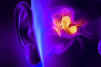
Image: A new way of imaging the middle ear uses infrared light (Photo courtesy of MIT).
The deeper tissue penetration of SWIR light not only allows better structure visualization, but also holds the potential of middle ear fluid detection, which has significant implications for diagnosing otitis media, the overdiagnosis of which is a primary factor in increased antibiotic resistance. The researchers found that middle ear fluid shows strong light absorption between 1,400 and 1,550 nm, enabling facile detection in a model using the SWIR otoscope. The study was published on August 22, 2016, in the Proceedings of the National Academy of Sciences (PNAS).
“The one clear diagnostic sign of an infection in the ear is a buildup of fluid behind the eardrum; but the view through a conventional otoscope can't penetrate deeply enough into the tissues to reveal such buildups,” said study co-author Jessica Carr, MSc, an MIT doctoral student. “A lot of times, it's a fifty-fifty guess as to whether there is fluid there. One of the limitations of the existing technology is that you can't see through the eardrum, so you can't easily see the fluid. But the eardrum basically becomes transparent to our device.”
Visualizing structures deep inside opaque biological tissues is one of the central challenges in biomedical imaging. Optical imaging with visible light provides high resolution and sensitivity; however, scattering and absorption of light by tissue limits the imaging depth to superficial features. SWIR shares many of the advantages of visible imaging, but light scattering in tissue is reduced, providing sufficient optical penetration depth to noninvasively interrogate subsurface tissue features.
Related Links:
Massachusetts Institute of Technology
University of Connecticut Health Center


