Cone Beam CT Imaging Could Help Detect Joint Osteoarthritis Earlier
By MedImaging International staff writers
Posted on 16 Aug 2016
Combining cone beam computed tomography (CBCT) with complementary metal-oxide semiconductor (CMOS) sensors could detect early stage osteoarthritis (OA).Posted on 16 Aug 2016
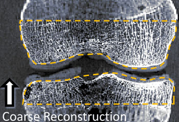
Developed by researchers at Johns Hopkins University (JHU, Baltimore, MD, USA), the portable CBCT/CMOS system could be used in doctors' offices, helping to detect OA with a radiation dose lower than that of standard computed tomography (CT). And since the CMOS sensor provides very high spatial resolution, it allows detection of finer details than standard CT, greatly enhancing doctors’ ability to assess trabecular bone. According to the researchers, the imaging system also could help with early identification of osteoporosis.

Image: Imaging reconstruction of a joint using CBCT/CMOS (Photo courtesy of JHU).
The researchers used the CBCT/CMOS system to image the hand of a human cadaver, as well as pelvic bone biopsy samples from 25 living women. They determined that the system provided results that correlate with micro-CT with an amorphous silicon (aSi) flat-panel detector (FPD), which is the gold standard for assessing trabecular bone. The preliminary results were presented at the 58th annual meeting of the American Association of Physicists in Medicine (AAPM), held during July-August 2016 in Washington (DC, USA).
“The technology we are developing allows us to see very fine detail in the mesh-like microstructure of bone – known as trabecular bone – which currently can't be assessed in patients,” said senior author medical physicist Wojciech Zbijewski, PhD, of the JHU department of biomedical engineering. “There is growing evidence that early stages of osteoarthritis involve changes in trabecular bone, so if we can detect such change, a patient could potentially avoid painful knee replacement surgery by getting treatment before the cartilage was irreversibly damaged.”
Currently, joints are usually assessed by x-ray or magnetic resonance imaging (MRI), which show if the cartilage lining the joint has been damaged. In most cases, by the time a patient has pain, the damage to the joint is irreversible, and the only solution is to surgically replace that joint. Joint replacement is therefore a common procedure, with 719,000 knee replacements and 332,000 hip replacements performed every year in the United States alone, according to the Centers for Disease and Prevention (CDC; Atlanta, GA, USA).
Related Links:
Johns Hopkins University


