Brain Imaging Technology Could Help Study Parkinson’s Disease
By MedImaging International staff writers
Posted on 27 Dec 2015
A portable imaging device has identified differences in brain activation patterns associated with postural stability in people with Parkinsonian syndromes, according to a new study. Posted on 27 Dec 2015
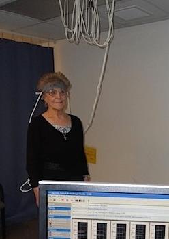
Researchers at Drexel University (Philadelphia, PA, USA) and Yeshiva University (YU; New York, NY, USA) used a portable functional near infrared spectroscopy (fNIRs) device as a direct online cortical probe to compare neural activation patterns in the prefrontal cortex, postural stability, and their respective interactions, in 269 older adults (mean 76 years, 56% women) without dementia. Of these, 26 patients had Parkinsonian syndromes; 117 had mild Parkinsonian signs; and 127 were healthy older adults who served as controls.

Brain Imaging Technology Could Help Study Parkinson’s Disease
The participants were asked to stand upright and count silently for ten seconds while changes in oxygenated hemoglobin levels over prefrontal cortex were measured using fNIRs. The researchers simultaneously evaluated postural stability with center of pressure velocity data recorded on an instrumented walkway. The results showed that when compared to healthy controls and patients with mild Parkinsonian signs, the patients with Parkinsonian syndromes demonstrated significantly higher prefrontal oxygenation levels to maintain postural stability. The study was published in the November 6, 2015, issue of Brain Research.
“Postural instability is a major risk factor for older adults. This initial study allowed us to measure brain activity in real time, in a realistic setting. It shows that there are indeed differences in the prefrontal cortex of healthy and Parkinsonian syndrome patients,” said study coauthor biomedical engineer Meltem Izzetoglu, PhD, of Drexel University. “If we can monitor the cognitive component of staying balanced, then this could eventually lead to better treatment options for people with Parkinsonian syndromes or even Parkinson's disease.”
The prefrontal cortex is the brain region considered to be in charge of the orchestration of thoughts and actions in accordance with internal goals, and is responsible for higher-level processing, such as memory, attention, problem solving, and decision-making. When a person is learning a new skill, for instance, neural activity is greater in this region. Increasing evidence shows that in Parkinson‘s disease, profound dopamine depletion not only occurs in the striatum of the brain, but also in the prefrontal cortex, and this may be associated with cognitive and motor deficits.
Related Links:
Drexel University
Yeshiva University


