Consumer Sensors Could Drastically Reduce Cost of Biomedical Imaging
By MedImaging International staff writers
Posted on 07 Dec 2015
A new study reveals how mathematical modeling could enable biomedical imaging systems to use a simple depth sensor to approximate the measurements of expensive lab equipment. Posted on 07 Dec 2015
Developed by researchers at the Massachusetts Institute of Technology (MIT; Cambridge, MA, USA), the imaging system is based on a technique called fluorescence lifetime imaging (FLI), which also has applications in DNA sequencing and cancer diagnosis. And while traditional FLI uses light bursts in the picosecond range, and FLI time in biomedical imaging is in nanosecond range, off-the-shelf depth sensors (like the Microsoft Kinect) use light bursts that last tens of nanoseconds, far too coarse for to biomedical FLI.

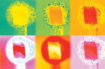
Image: Phase information contained in six of the 50 light frequencies analyzed by the system (Photo courtesy of MIT).
The MIT researchers, however, were able to extract additional information from the fluorescent light signal by subjecting it to a Fourier transform, breaking it into its constituent frequencies. The optical signal returning from a biological sample is the sum of 50 different frequencies, which enabled the researchers to recover information from FLI bursts shorter than actual duration of the emitted burst of light. They did so by calculating each phase shift using a mathematical model of the overlapping intensity profiles of both the reflected and re-emitted light.
Once the deduced intensity profile of the reflected light is known, the distance between the emitter and the sample can be calculated. So unlike conventional FLI, the researchers' approach does not require calibration. To test the model, the researchers used commercial depth sensors that cost USD 100 with arrays of roughly 20,000 light detectors each. They found that while the setup does not afford the image resolution of existing FLI microscopes, it was good enough for some purposes to replace USD 100,000 lab equipment with denser arrays. The study was published in the November 20, 2015, issue of Optica.
“The theme of our work is to take the electronic and optical precision of this big expensive microscope and replace it with sophistication in mathematical modeling,” said lead author Ayush Bhandari, MSc, a graduate student at the MIT Media Lab. “We show that you can use something in consumer imaging, like the Microsoft Kinect, to do bioimaging in much the same way that the microscope is doing.”
FLI measures that interval—the lifetime of the fluorescence event—in a biological sample treated with a fluorescent dye to reveal information about the sample's chemical composition, since for any given fluorophore, interactions with other chemicals will shorten the interval between the absorption and emission of light in a predictable way.
Related Links:
Massachusetts Institute of Technology


