Handheld Probe Generates Precise Images Without Bulky, Costly Instruments
By MedImaging International staff writers
Posted on 17 Nov 2014
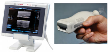
A new handheld probe developed by university and industry researchers in the Netherlands and France could give clinicians useful new imaging capabilities that fit in the palms of their hands. A technology that once filled up a whole lab bench has been shrunken down to a computer screen and a small probe approximately the size of a stapler. Posted on 17 Nov 2014
The new device combines two imaging modalities: ultrasound and photoacoustics. Ultrasound is a well-accepted technology that analyzes how sound pulses echo off internal body parts. It is suitable for revealing anatomic structures and is, perhaps most familiarly, used to image a developing fetus in a mother’s womb. The device was described October 20, 2014, in a paper published in The Optical Society’s (OSA) open-access journal Optics Express.
Photoacoustics is a relatively new imaging technique, still making its way toward widespread clinical applications. In photoacoustic imaging, short pulses of light heat up internal tissue. The slight temperature change leads to an alteration in pressure, which then produces a wave of ultrasound that can be examined to show data about the body’s internal workings. Since this technique ultimately produces ultrasound waves as well, existing technology can be used to analyze and display the images.
The advantage of photoacoustics is that it can reveal important medical information that other imaging techniques cannot, including the presence of molecules such as hemoglobin and melanin and the submillimeter structure of complexes of blood vessels several centimeters beneath the skin. When combined with spectroscopic measurements, photoacoustics can also quantify hemoglobin oxygen saturation within single vessels, providing metabolic information that could be helpful for monitoring tumor progression.
However, in spite of these benefits, the cost and size of most photoacoustic systems restrict their widespread use, according to Dr. Khalid Daoudi, a researcher in the biomedical photonic imaging group at the University of Twente (Enschede, the Netherlands). Most systems on the market require expensive and cumbersome lasers that make the systems unfeasible for point-of-care (POC) diagnostics. “Our research aimed to break through these hindering factors,” Dr. Daoudi said.
The project began as collaboration between the University of Twente and three European companies: Esaote Europe (Maastricht, The Netherlands), a maker of medical diagnostic systems, Quantel (Newbury, Berkshire, UK), a maker of solid-state lasers, and SILIOS Technologies (Peynier, France), a developer of optical components.
The investigators’ major development, which allowed them to drastically shrink the system, was the design of an ultra-compact laser based on an effective and inexpensive laser diode. By stacking multiple diodes to raise the power and carefully designing optical elements to shape the laser beam, the scientists were able to generate laser pulses with energies higher than had ever been achieved before with diode technology.
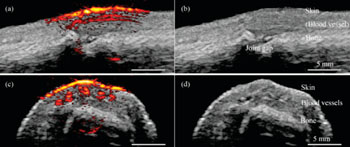
Diode lasers can also provide many laser pulses per second, which in turn allows real time imaging, another advantage of the new system, Dr. Daoudi noted. The researchers evaluated the imaging performance of the system in different types of phantoms and in a healthy human finger joint. The new compact probe and imaging system can be easily transported between rooms in a clinical environment, an appealing feature for future commercialization, the researchers reported.
The researchers are now working with a European consortium of industrial and academic partners to take the next phase, moving the research to commercialization. The current system operates at a single wavelength in the near infrared, but the team has plans to expand the design to multi-wavelength imaging. “Some applications targeted are rheumatoid arthritis in finger joints, oncology, cardiovascular disease and burn wounds,” Dr. Daoudi said.
Related Links:
University of Twente
Esaote Europe
Quantel


