“Nanojuice” and Photoacoustic Tomography Combo Developed to Enhance Visualization of the Gatrointestinal Tract
By MedImaging International staff writers
Posted on 24 Jul 2014
Located deep in the human gut, the small intestine is not an organ that is easy to image and examine. Magnetic resonance imaging (MRI), X-rays, and ultrasound scans provide images but each has limitations. However, researchers are developing a new imaging technique involving nanoparticles suspended in liquid to form “nanojuice” that patients would drink. Upon reaching the small intestine, clinicians would hit the nanoparticles with a harmless laser light, providing an unprecedented noninvasive, real-time view of the organ. Posted on 24 Jul 2014
Described July 6, 2014, in the journal Nature Nanotechnology, this new advance could help clinicians better identify, determine, and treat gastrointestinal (GI) disorders. “Conventional imaging methods show the organ and blockages, but this method allows you to see how the small intestine operates in real time,” said corresponding author Jonathan Lovell, PhD, University at Buffalo (UB; NY, USA) assistant professor of biomedical engineering. “Better imaging will improve our understanding of these diseases and allow doctors to more effectively care for people suffering from them.”

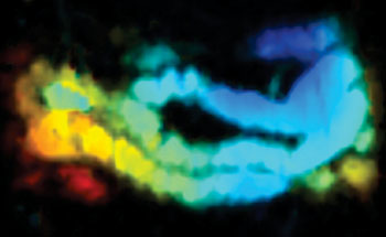
Image: The combination of “nanojuice” and photoacoustic tomography (PAT) illuminates the intestine of a mouse (Photo courtesy of Jonathan Lovell, University at Buffalo).
The average human small intestine is approximately 7-meters-long and 2.5-cm-thick. Squeezed in between the stomach and large intestine, it is where much of the digestion and absorption of food takes place. It is also where symptoms of irritable bowel syndrome, celiac disease, Crohn’s disease and other gastrointestinal disorders occur. To assess the organ, doctors typically require patients to drink a thick, chalky liquid called barium. Radiologists then use X-rays, MRI, and ultrasound scanning to assess the organ, but these techniques are limited with respect to safety, accessibility and lack of adequate contrast, respectively.
Furthermore, none are highly effective at providing real-time imaging of movement such as peristalsis, which is the contraction of muscles that moves food through the small intestine. Dysfunction of these movements may be linked to the previously mentioned illnesses, as well as side effects of thyroid disorders, diabetes and Parkinson’s disease. Dr. Lovell and a team of researchers worked with a class of dyes called naphthalcyanines. These small molecules absorb large portions of light in the near-infrared spectrum, which is the ideal range for biologic contrast agents. They are unsuitable for the human body, however, because they do not disperse in liquid and they can be absorbed from the intestine into the blood stream.
To address these hurdles, the researchers formed nanoparticles called “nanonaps” that contain the colorful dye molecules and added the abilities to disperse in liquid and move safely through the intestine. In laboratory research performed with mice, the researchers administered the nanojuice orally. They then used photoacoustic tomography (PAT), which is pulsed laser lights that generate pressure waves that when measured provide a real-time and more nuanced view of the small intestine.
The researchers next plan to continue to modify the technique for human trials, and move into other areas of the GI tract.
Related Links:
University at Buffalo


