Innovative Dye Improves Diagnostic Imaging
By MedImaging International staff writers
Posted on 22 May 2013
Imaging technology is becoming more vital in hospitals worldwide, from magnetic resonance imaging (MRI) scanners to microscopes, whether for the diagnosis of disorders or for research into new cures. Imaging technology requires dyes or contrast agents of some sort. Current contrast agents and dyes are costly, difficult to work with, and far from ideal. Now, Danish chemists have discovered a new dye that they report is superior to any of the dyes currently available. Posted on 22 May 2013
Drs. Thomas Just Sørensen and Bo Wegge Laursen are chemists at the University of Copenhagen (Denmark). In a series of published articles, they have shown that the aza-oxa-trangulenium dyes have the potential to outclass all fluorescent dyes currently used in imaging. “Our dyes are 10 times better, far cheaper, and easier to use. The latter I believe, will lead to expanded opportunities and broadened use, by physicians and researchers in developing countries, for example,” stated Dr. Sørensen.

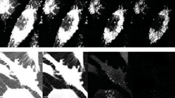
Image: Human breast cancer cells stained with triangulenium and state of art dye. Picture series shot over 30 nanoseconds. Triangulenium emits light up to 100 nanoseconds (Photo courtesy of the University of Copenhagen).
One of the key challenges, interestingly, when capturing images of cells and organs, is to avoid noise. The agents that make it possible to see microscopic biologic structures are luminescent, but then, so is tissue. Consequently, the contrast agent’s light risks being overpowered by “light noise.” Tissue becomes luminescent when exposed to light, similar to the hands of a clock. Tissue and other organic structures luminesce, or lights up, for 10 nanoseconds after exposure to light. The light-life of an ordinary dye is the same—10 nanoseconds. However, triangulenium dyes produce light for an entire 100 nanoseconds.
The long life of the triangulenium dyes means that an image can be generated without background noise. Furthermore, the extra 90 nanoseconds opens the potential of filming living images of the processes occurring within cells, for instance, when a drug attacks an illness.
Medical image analysis departments currently spend a lot of time staining samples, because all samples must be treated with two agents. The use of triangulenium dyes necessitates only one dye. Furthermore, in contrast with typical dyes, no specialized equipment is needed to see the dyes in tissue samples. A lens from a pair of polarized sunglasses and a simple microscope are all that are required.
When one compares the benefits of triangulenium dyes against the three million Danish kroner per gram price tag of traditional dyes (USD 500.000) one would expect that the new dye would immediately out-compete its predecessors. However, up to now, Drs. Sørensen and Laursen have had to give their dye away. “I know that our dye is better, but biologists and physicians don’t. Therefore, we are giving the dye away to anyone that wants to perform a comparison test. Someone who needs to assess the health of sick people wouldn’t dare to rely on an untested substance. Only when several researchers have shown triangulenium dyes to perform just as effectively as its predecessors can we hope for our substance to become more widely adopted,” concluded Dr. Sørensen.
The latest findings on the agent were published in May 2013 issue of the journal Analytical and Bioanalytical Chemistry.
Related Links:
University of Copenhagen


