Brightest X-Ray Machine Probes Molecules
By MedImaging International staff writers
Posted on 17 Jun 2010
The brightest X-ray machine in the world has been designed for the energies at which it operates--with photon energies in the "hard X-ray” region and very high beam intensities of 1,018 W/cm2. At these energies, the machine can serve as an extraordinary microscope for viewing matter at the scale of atoms, and biologists, chemists, and physicists have been eager to do precisely that. The technology also acts like a knife since it can peel electrons away from the parent atoms and molecules, even those huddling very close to the nucleus.Posted on 17 Jun 2010
The Stanford [University] Linear Accelerator Center (SLAC; Stanford, CA, USA), long the preserve of particle physics, is also a major laboratory for conducting experiments in fields like biology and medicine. The electron acceleration equipment has been adapted over the past few years to create something known as the Linac Coherent Light Source (LCLS), which produces short X-ray pulses millions of times brighter than those currently created by other instruments.

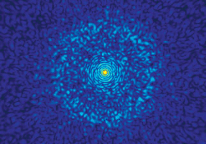
Image: Snapshot from a Livermore simulation predicts the diffraction pattern that will occur when the Linac Coherent Light Source shoots an X-ray beam at a nanolipoprotein particle (NLP). Atomic bonds appear in the pattern’s outer rings. The yellow–red spot in the middle indicates where 95 percent of the beam traveled through the NLP to the machine’s beam stop (Photo courtesy of Lawrence Livermore National Laboratory).
Becoming operational in the fall of 2009, the first experimental results from the LCLS are starting to appear at scientific meetings. Dr. Li Fang of Western Michigan University (Kalamazoo, MI, USA) reported on May 21, 2010, at the 2010 Conference on Lasers and Electro-Optics/Quantum Electronics and Laser Science Conference (CLEO/QELS), held in San Jose, CA, USA, on how the powerful LCLS X-rays can be used to strip electrons away from a nitrogen molecule. He noted that in the extreme case, nitrogen atoms were detected from which all of the electrons had been removed. This causes the molecule to quickly dissociate. The plucked electrons, which nearby detectors can spot and measure, allow researchers to calculate the binding energy within the original molecule.
In future studies, more such measurements should give investigators a more accurate assessment of large molecules, in particular, biomolecules.
At the 2010 CLEO/QELS, researchers from around the world presented the latest breakthroughs in electro-optics, innovative developments in laser science, and commercial applications in photonics.
Related Links:
Stanford Linear Accelerator Center
Western Michigan University


 Guided Devices.jpg)


