Deep Learning Enables Fast 3D Brain MRI at 0.055 Tesla
Posted on 27 Sep 2023
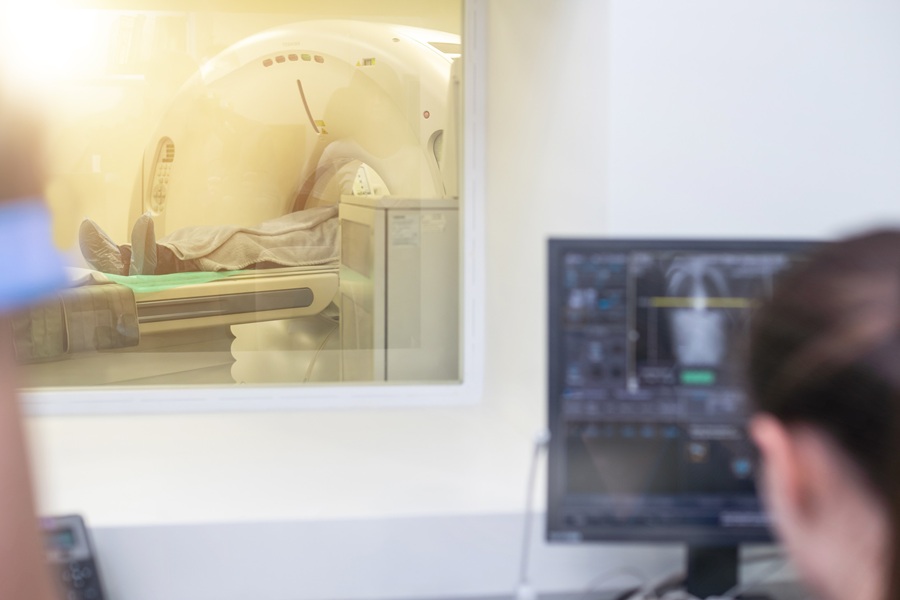
In the past few years, there has been significant work on developing portable ultralow-field magnetic resonance imaging (MRI) machines. These are designed to be affordable, easy to use without special shielding, and good for point-of-care settings. However, their image quality has not been up to the mark, and scans also take a long time. Deep learning is changing the game and has been effective in various high-field MR image reconstruction tasks, including artifact, denoising, and reconstruction. Now, researchers have proposed a fast acquisition and deep learning reconstruction framework to speed up brain MRI at 0.055 tesla.
In a new study aiming to advance brain ultralow-field MRI for speed and quality using deep learning, researchers at The University of Hong Kong (Hong Kong, PRC) successfully achieved fast 3D brain MRI at 0.055 T. This was done by directly combining the accelerated MR data acquisition and the deep learning 3D image formation that reconstructs images from incomplete 3D k-space data with super-resolution. The acquisition comprises a single average 3D encoding with 2D partial Fourier sampling, cutting down the scan time of T1- and T2-weighted imaging protocols to 2.5 and 3.2 minutes, respectively.
")
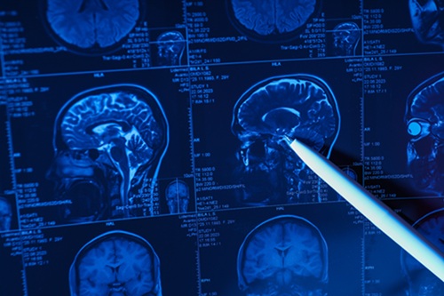
The 3D deep learning system takes advantage of the homogeneous brain anatomy available from high-field MRIs to enhance image quality. It minimizes errors and noise while boosting the spatial resolution to a synthetic 1.5-mm isotropic resolution. The new approach successfully overcomes the limitations of low signal strength, allowing for the detailed reconstruction of fine anatomical features. These features are consistent both within individual subjects and across different imaging protocols. This new method opens the door to fast and high-quality whole-brain MRIs at a magnetic field strength of 0.055 tesla, offering a variety of potential applications in biomedicine.
Related Links:
The University of Hong Kong