Computer Program Bests Radiologists at Analyzing Brain MRI
By MedImaging International staff writers
Posted on 27 Sep 2016
A new study that matched two physicians against a computer algorithm in analysis of magnetic resonance imaging (MRI) brain scans found the program was nearly twice as accurate.Posted on 27 Sep 2016
Researchers at Case Western Reserve University (CWRU; Cleveland, OH, USA) and the University of Texas Southwestern Medical Center (Dallas, TX, USA) conducted a study to determine the feasibility of using computer-extracted texture features to differentiate between radiation necrosis and recurrent brain tumors on post-radiochemotherapy MRI scans. In all, 58 patient scans were used, with 43 forming the training cohort for the algorithm and 15 forming the test cohort.

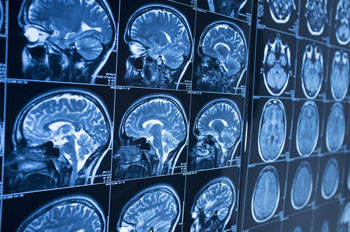
Image: A computer program beats radiologists in MRI analysis (Photo courtesy of CWRU).
A set of radiomic features was extracted for every lesion on each MRI sequence - gadolinium T1WI, T2WI, and FLAIR. Feature selection was used to identify the top five most discriminating features for every MRI sequence on the training cohort. These features were then evaluated on the test cohort by a support vector machine classifier. The classification performance was compared against diagnostic reads by two expert neuroradiologists, who had access to the same MRI sequences as the classifier. Finally, clinical histologic findings were confirmed by an experienced neuropathologist.
The results revealed that in the direct comparison, one neuroradiologist diagnosed seven patients correctly, and the second physician correctly diagnosed eight patients. The computer program, on the other hand, was correct on 12 of the 15 MRI scans. The researchers are now seeking to validate the algorithms' accuracy using a much larger collection of images from across different sites, so that it could eventually be used as a decision support tool to assist neuroradiologists in improving their confidence in identifying a suspicious lesion. The study was published on September 15, 2016, in the American Journal of Neuroradiology.
“One of the biggest challenges with the evaluation of brain tumor treatment is distinguishing between the confounding effects of radiation and cancer recurrence; on an MRI, they look very similar,” said lead author biomedical engineer Pallavi Tiwari, PhD, of CWRU. “What the algorithms see that the radiologists don't are the subtle differences in quantitative measurements of tumor heterogeneity and breakdown in microarchitecture on MRI, which are higher for tumor recurrence.”
“While the physicians use the intensity of pixels on MRI scans as a guide, the computer looks at the edges of each pixel; if the edges all point to the same direction, the architecture is preserved,” added senior author professor of biomedical engineering Anant Madabhushi, PHD, director of the CWRU Center of Computational Imaging and Personalized Diagnostics. “If they point in different directions, the architecture is disrupted—the entropy, or disorder, and heterogeneity are higher.”
In a recent competition at the 2016 International Symposium of Biomedical Imaging (ISBI), held during April in Prague (Czech Republic), a machine-learning computer algorithm that was trained to recognize breast cancer metastasis in lymph nodes identified correctly 92% percent of the time, nearly matching the 96% success rate of a human pathologist.
Related Links:
Case Western Reserve University
University of Texas Southwestern Medical Center


.jpg)