Algorithm Outperforms Radiologists in Detecting Pneumonia on X-Rays
By MedImaging International staff writers
Posted on 21 Nov 2017
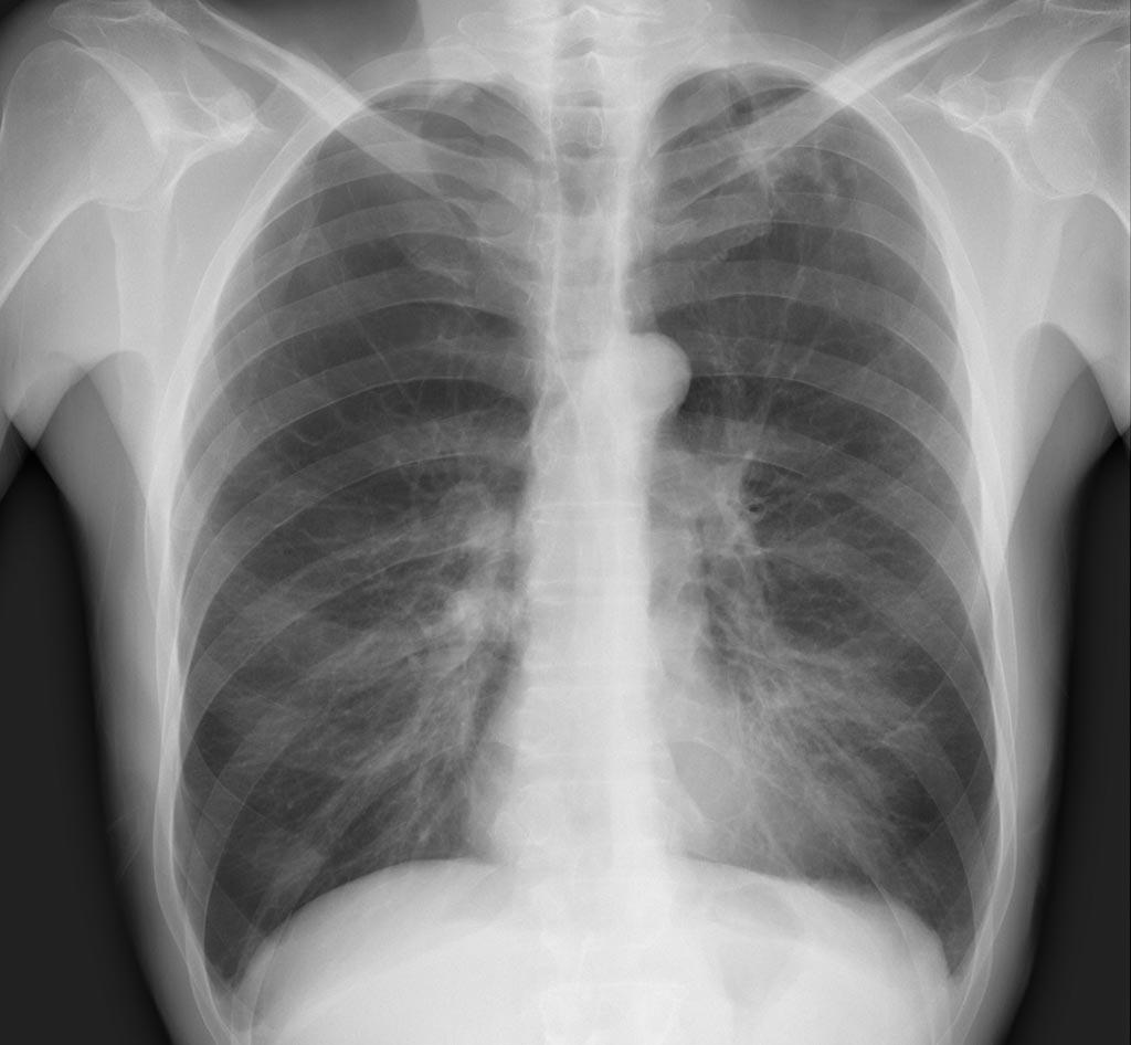
A deep learning algorithm developed by researchers from the Stanford University (Stanford, CA, USA) that evaluates chest X-rays for signs of disease has outperformed expert radiologists at diagnosing pneumonia in just over a month of its development. A paper about the algorithm named CheXNet, which can diagnose up to 14 types of medical conditions, was published November 14 on the open-access, scientific preprint website arXiv.Posted on 21 Nov 2017
Soon after the National Institutes of Health Clinical Center recently released a public dataset containing 112,120 frontal-view chest X-ray images labeled with up to 14 possible pathologies, the Machine Learning Group at Stanford began developing an algorithm that could automatically diagnose the pathologies. Meanwhile, four Stanford radiologists independently annotated 420 of the images for possible indications of pneumonia. Within a week the researchers had developed an algorithm that diagnosed 10 of the pathologies labeled in the X-rays more accurately than the previous state-of-the-art results. In just over a month, CheXNet could beat these standards in all 14 identification tasks and also outperformed the four individual Stanford radiologists in pneumonia diagnoses.

Image: The algorithm CheXNet can diagnose up to 14 types of medical conditions, including pneumonia (Photo courtesy of Stanford ML Group).
The Stanford researchers have also developed a computer-based tool that produces what appears to be a heat map of chest X-rays, although instead of representing temperature, the colors of these maps represent the areas determined by the algorithm as the ones most likely to represent pneumonia. The tool could help reduce the amount of missed pneumonia cases and significantly accelerate the workflow of radiologists by indicating where to look first, resulting in faster diagnoses for the sickest patients.
“We plan to continue building and improving upon medical algorithms that can automatically detect abnormalities and we hope to make high-quality, anonymized medical datasets publicly available for others to work on similar problems,” said Jeremy Irvin, a graduate student in the Machine Learning group and co-lead author of the paper. “There is massive potential for machine learning to improve the current health care system, and we want to continue to be at the forefront of innovation in the field.”
Related Links:
Stanford University


