AI-Powered Mammograms Predict Cardiovascular Risk
Posted on 21 Mar 2025
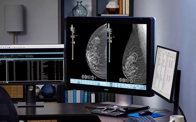
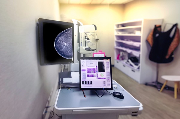
The U.S. Centers for Disease Control and Prevention recommends that women in middle age and older undergo a mammogram, which is an X-ray of the breast, every one or two years to screen for breast cancer. In the United States, approximately 40 million mammograms are performed annually. A new study, presented at the American College of Cardiology’s Annual Scientific Session, suggests that mammograms, when combined with artificial intelligence (AI) models, can reveal much more than just cancer. The findings show that these essential screening tools can also be used to assess the amount of calcium buildup in the arteries within breast tissue, which is an important indicator of cardiovascular health.
Heart disease is the leading cause of death in the United States, but it remains underdiagnosed in women, and awareness is still lacking. The buildup of calcium in blood vessels is a sign of cardiovascular damage linked to early-stage heart disease or aging. Studies have demonstrated that women with calcium deposits in their arteries have a 51% higher risk of heart disease and stroke. Although breast artery calcifications can be detected in the images, radiologists typically do not quantify or report this information to women or their healthcare providers. In this new study, researchers from Emory University (Atlanta, GA, USA) and Mayo Clinic (Rochester, MN, USA) employed an AI image analysis technique, previously unused in mammograms, to show how AI can assist by automatically analyzing breast arterial calcification and converting the findings into a cardiovascular risk score.
")
According to the researchers, AI-enabled mammogram screening tools could help identify more women with early signs of cardiovascular disease, maximizing the use of the routine screening tests many women already undergo. To develop the screening tool, researchers trained a deep-learning AI model to segment calcified blood vessels in mammogram images—these calcifications appear as bright spots on X-rays—and compute the future risk of cardiovascular events using data obtained from electronic health records. This segmentation method sets this model apart from previous AI models designed for analyzing breast artery calcifications. The model was also enhanced by the use of a large dataset for training and testing, which included images and health records from more than 56,000 patients who had mammograms at Emory Healthcare between 2013 and 2020, along with at least five years of follow-up electronic health records.
The results of the study showed that the new AI model was effective in classifying patients' cardiovascular risk as low, moderate, or severe based on mammogram images. After evaluating the risk of dying from any cause or experiencing an acute heart attack, stroke, or heart failure within two and five years, the model revealed that the rate of serious cardiovascular events increased as the level of breast arterial calcification rose in two of the three age groups—women under 60 and those between 60 and 80—but not in those over 80. This suggests that the tool is particularly valuable for detecting heart disease risk early in younger women, who can benefit most from early interventions. The study also found that women with the highest levels of breast arterial calcification (above 40 mm²) had a significantly lower five-year event-free survival rate compared to those with lower levels (below 10 mm²).
Specifically, 86.4% of those with severe breast arterial calcification survived for five years, compared to 95.3% of those with little or no calcification. This translates to approximately 2.8 times the risk of death within five years for patients with severe breast arterial calcification compared to those with minimal calcification. The AI model is not yet available for use, but if it passes external validation and gains approval from the U.S. Food and Drug Administration, it could be made commercially available for other healthcare systems to incorporate into routine mammogram processing and follow-up care. The researchers also plan to explore how similar AI models could be applied to assess biomarkers for other conditions, such as peripheral artery disease and kidney disease, that might be identified through mammograms.
“We see an opportunity for women to get screened for cancer and also additionally get a cardiovascular screen from their mammograms,” said Theo Dapamede, MD, PhD, a postdoctoral fellow at Emory University in Atlanta and the study’s lead author. “Our study showed that breast arterial calcification is a good predictor for cardiovascular disease, especially in patients younger than age 60. If we are able to screen and identify these patients early, we can refer them to a cardiologist for further risk assessment.”
Related Links:
Emory University
Mayo Clinic