New SPECT/CT Technique Could Change Imaging Practices and Increase Patient Access
Posted on 18 Apr 2024
The development of lead-212 (212Pb)-PSMA–based targeted alpha therapy (TAT) is garnering significant interest in treating patients with metastatic castration-resistant prostate cancer. The imaging of 212Pb, however, presents challenges due to the high-energy gamma rays emitted by the isotope, which produce considerable scatter. Now, a novel SPECT/CT acquisition method utilizing 212Pb can conveniently detect radiopharmaceutical biodistribution in prostate cancer patients, facilitating more tailored treatments. Researchers have published the first-in-human images from this new imaging technique which could revolutionize practices and broaden patient access globally.
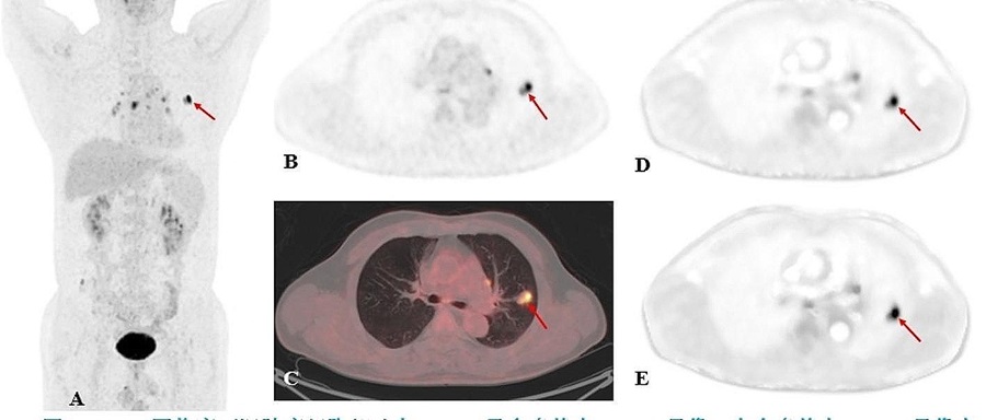
In this pioneering study conducted by researchers at AdvanCell (Sydney, Australia), a dose of 60 MBq of 212Pb-ADVC001 was administered to a 73-year-old male patient diagnosed with metastatic castration-resistant prostate cancer. The SPECT/CT imaging was performed at intervals of 1.5, 5, 20, and 28 hours following the infusion. The representative 212Pb SPECT/CT images displayed rapid initial tumor uptake of 212Pb-ADVC001, consistent with the tumor burden identified in pretreatment 18F-DCFPyl PET/CT scans. The scans conducted at 20 hours post-infusion continued to show significant tumor uptake, although the observable counts were lower due to the decay of 212Pb.
")
Going forward, this innovative imaging technique is poised to enhance the drug development process by ensuring the efficacy of agents before they are escalated to larger-scale trials. Moreover, the capability to image 212Pb using standard SPECT cameras within a relatively short timeframe confirms its utility as a true theranostic alpha-emitter. This advancement holds great promise for optimizing patient selection for targeted alpha therapies.
“The ability to acquire imaging of an alpha-emitter with a standard SPECT camera and standard collimator within a convenient acquisition time for the patient could provide more precision in how we treat patients with prostate cancer, and patients with other cancers, in the future,” said Stephen Rose, PhD, head of Translational Medicine and Clinical Science at AdvanCell. “Access to PET imaging is a bottleneck, in the United States and globally. SPECT cameras are more widely available and may address this critical issue, as SPECT imaging can be used for patient selection, therapy decision making, and guiding adaptive dosing strategies based on changes of target expression and tumor volume during treatment.”