Digital Radiography Table Advances Clinical Diagnostics
By MedImaging International staff writers
Posted on 24 Aug 2016
A new digital radiographic table system offers high-performance digital projection radiography with features that supports productivity, user-operability, patient comfort, and safety.Posted on 24 Aug 2016
The RADspeed Pro Edge Digital Radiography package of advanced applications includes tomosynthesis, which allows the user to obtain multiple digital cross section images from a single linear tomography scan. The raw scan data can then be used to reconstruct cross sectional images from any height, as many times as desired. Furthermore, it offers the flexibility of scanning patients while applying loads in the standing position, in the supine position on the table, or with elbows or knees bent. A metal artifact inhibition feature helps evaluate bones near metal objects, such as used for checking bone union status after orthopedic surgery.
.")
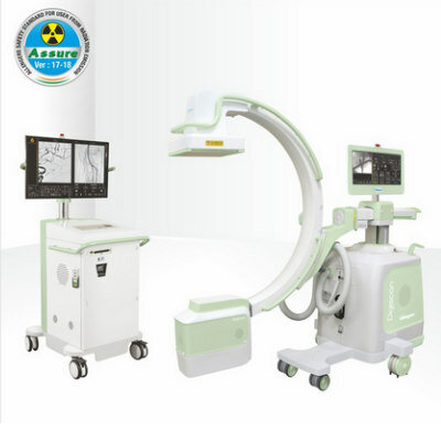
Image: The RADspeed Pro Edge Digital Radiography package (Photo courtesy of Shimadzu).
Another innovative feature is dual energy subtraction, which utilizes the difference in X-ray absorption levels of bones and soft tissues to generate separate images of each, in addition to plain radiographic images, by successively applying high- and low-voltage exposures and then using an algorithmic process to separate them. This enables rendering of nodes obscured behind ribs in soft-tissue images of the chest area or calcifications in bone images, which are especially useful for diagnoses of the chest area, such as with lung cancer.
The newly developed Bucky table is able to obtain 120 cm long view images in the supine position and 160 cm standing long view images, helping to visualize the entire spine or entire lower extremities, even for large patients or patients with difficulty in standing. Automatic linking of the settings made on the X-ray tube with the Bucky table or Bucky stand allows the system to perform subsequent automatic stitching of the radiographs to generate an anatomically coherent image.
Innovative flat panel detectors (FPD) in various cassette formats offer maximum efficiency in clinical practice; large format, 43x43 cm and 34x43 cm detectors for general radiographic examinations are available, equipped with a Cesium Iodide (CsI) or Gadolinium oxysulfide (GOS) scintillator. Additionally, a smaller sized FPD with an imaging area of 24x30 cm allows free positioning on the X-ray table, such as for dedicated orthopedic examinations. The RADspeed Pro Edge Digital Radiography package is a product of Shimadzu (Kyoto, Japan).
Related Links:
Shimadzu

Gold Member
Solid State Kv/Dose Multi-Sensor
AGMS-DM+
C-Arm with FPD
Digiscan V20 / V30

New
Digital Radiography Generator
meX+20BT lite

New
X-Ray Detector
FDR-D-EVO III

.jpg)